Why does cellulite appear?
The main causes of cellulite:
- Increase in the volume of subcutaneous fat.
- Impaired blood supply in a specific area.
An example of the first reason. The girl begins to eat improperly; her body receives an excess amount of easily digestible nutrients, which are actively stored in fat cells. When the intake of active substances exceeds their consumption, this leads to the fact that these substances begin to accumulate in fat cells. This leads to the fact that fat cells begin to increase in volume - as a result, the subcutaneous fat layer increases. Enlarged fat cells begin to compress everything around them - primarily the vessels that feed these same cells. Accordingly, the blood supply to the subcutaneous fat tissue deteriorates, the flow of oxygen decreases, and the development of connective fibrous tissue begins, which is the main clinical sign of cellulite.
An example of the second reason is sedentary work. A girl can be thin and eat right, but sedentary work leads to compression of the soft tissues in the back of the thigh, which leads to a disruption in blood supply and, as a result, oxygen supply. The mechanics of the process are identical to those described above, only the reason differs. This is why “orange peel” often appears in absolutely thin girls.
Gender matters
But why does this happen in women and not in men? The reason for this is that women have a distinctive structure of collagen fibers in the subcutaneous tissue. They are located perpendicular to the skin and parallel to each other. This leads to frequent fluid stagnation. The female hormone estrogen activates the process of “obesity” of subcutaneous cells.
Male collagen fibers intersect with each other, forming a strong cellular structure, which is much more difficult to stretch.
Let's make a small remark: it is impossible to visually determine male cellulite. But if a representative of the stronger half of humanity has significantly gained weight and is already in the second and third stages of obesity, he can still be diagnosed with cellulite. The thing is that with the development of female-type obesity in men, estrogens begin to accumulate in adipose tissue. So, with excess body weight, cellulite occurs in men.
Features of the appearance of cellulite in pregnant women
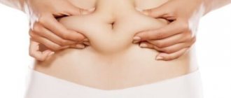
Women in an “interesting position” are at a particular risk of developing cellulite. The reason for this is constant changes in the body and stagnant processes in the cells of the subcutaneous fatty retina. After all, it is known that during pregnancy a woman begins to rapidly gain weight. Many body systems do not have time to cope with increased stress and “turn on” their defensive reaction. These include cells that stop their reproduction and begin to accumulate fluid, fats and toxins. This leads to the formation of an "orange peel".
Controversies in aesthetic medicine. 4. The Mystery of Cellulite
I. Kruglikov, Doctor of Physical and Mathematical Sciences, Wellkomet GmbH, Germany AESTHETIC MEDICINE, volume XII, No. 4, 2013
1. INTRODUCTION
Cellulite is a cosmetic problem that is known to affect more than 85% of all women over the age of 20 and which most often affects the lower extremities, in particular the gluteal-femoral region, and the abdomen. Treatment options are still very limited, and their results tend to be short-term effects.
The main reason for this is that the pathological physiology of cellulite still remains unclear, although many articles have been published on this topic in recent years. In accordance with various hypotheses, cellulite received many often contradictory names, which, according to their authors, were supposed to reflect its pathological physiology, including: nodular liposclerosis, edematous panniculopathy, fibrosclerotic panniculopathy, gynoid lipodystrophy, dermatomyoliposclerosis, etc.
In the pathophysiology of cellulite, the role of connective tissue remains a highly controversial issue. On the one hand, today there is virtually no doubt that collagen plays an important role in the formation of cellulite, on the other hand, its role is described very controversially.
Some hypotheses suggest that collagen structures in adipose tissue are weakened due to increased activity of matrix metalloproteinases (MMPs), responsible for the breakdown of collagen, and therefore are unable to withstand the pressure of fat trabeculae. From this we could conclude that it is necessary to consistently reduce collagen breakdown (due to inhibition of matrix metalloproteinases) to strengthen the collagen structure.
Other hypotheses proceed from the fact that the septa in cellulite are highly stretched, since they perform two main tasks at once - they compact adipocytes and provide structural support to the skin. With age, the septa become increasingly fibrotic, which increases skin tension, which leads to the formation of compartmental structures typical of cellulite. If we take this as a basis, the logical way out of the situation would be targeted cleavage of the fibrous structures of the connective tissue, which could be achieved by stimulating matrix metalloproteinases, that is, to carry out treatment absolutely opposite to that required based on the hypothesis of weakening of collagen structures.
A logical substantiation of the pathological physiology of cellulite is extremely necessary, first of all, for the development of a scientifically based and effective strategy for its treatment [1]. The theory should allow us to answer the following basic questions.
Why are the gluteal-femoral and abdominal areas most often affected by cellulite in women?
Why are there different stages of cellulite, which in severe cases can lead to an inflammatory condition (panniculitis)?
Why does cellulite respond to a greater or lesser extent to a variety of treatment methods, such as the use of heat, vacuum massage, elastic compression, ultrasound, radio frequency current, light, etc.? What is the non-specificity of these methods?
Why are the results of treatment short-lived, and why is the development of therapeutic resistance often observed when applying the sequence of procedures chosen for treatment?
Why are there significant differences in the occurrence of cellulite between representatives of the Mongoloid (Asian) and Caucasian (Caucasian) races?
This article describes a new pathological physiology of cellulite that may provide answers to the above questions.
2 FIBROSIS OF ADIPOSE TISSUE
Two important discoveries in recent years show that the role of collagen in adipose tissue has been largely underestimated. It was found that:
The extracellular matrix (Extracellular Matrix, ECM) of adipose tissue begins to change its structure from fibrillar to laminar as soon as adipocytes, due to the accumulation of triglycerides, enter the “expansion” phase [2]. In this case, not only a new structure of adipose tissue appears, but also the percentage of different types of collagen fibers included in its composition changes;
“adipotic” fibrosis develops in hypertrophied adipose tissue, which is a sign of metabolic changes in adipocytes [3, 4].
Fibrosis, a specific process occurring in hypertrophied adipose tissue [4], apparently plays an important role not only in the development of obesity, but also in the formation of cellulite. Fibrosis may be expressed differently in different types of adipose tissue, and this is likely due to the percentage of different types of collagen fibers they contain. The most important types of collagen in adipose tissue are I, III, IV, V and VI. Collagens of these types have a different structure: collagens of types I, III and V are fibrillar in their structure, IV – laminar (network-forming), and VI – microfibrillar (thread-like). All of these collagens are involved in fibrosis of adipose tissue.
Collagen fibers in hypertrophied subcutaneous adipose tissue are present in large quantities and are organized into dense bundles [3]. These fibrillar structures, which are thought to be primarily composed of collagen types I and III, penetrate the adipose tissue in different directions and constrict it. As fibrosis progresses, they can grow so large that they eventually form “island structures” in the tissue. The larger these structures, the more tension they create in the fatty tissue. This entire process can be considered as local scarring of adipose tissue, although histologically the fibrous structures in adipose and scar tissue may differ significantly. The similarities between scar tissue and fibrotic adipose tissue raise the question of whether similar treatments (eg, collagenase, elastocompression, etc.) can be prescribed for cellulite and scars. This possibility will be analyzed later in this article.
A different role in adipose tissue is played by collagens IV (the basement membrane around adipocytes, or the so-called basal lamina) and VI (microfibrils of the extracellular matrix) types. Both of them belong to non-fibrillar collagens and can be intertwined with each other [2]. Type VI collagen is found in large quantities in different types of adipose tissue, and its content increases markedly with increasing body mass index (BMI) [5, 6], and it also has the property of intensively binding hyaluronic acid [7]. Types IV and VI collagens together form structures that surround adipocytes and create pericellular fibrosis.
Fibrillar structures are often classically described as septa, and they are usually responsible for the appearance of cellulite skin. However, there are several important points that argue against such a simple interpretation. Firstly, the distance between septa in normal adipose tissue is within 1 mm, which, as we understand it, is much less than the distance between typical retractions on the surface of the skin with cellulite. Secondly, the resistance of septa to rupture is approximately 40 mJ/m2, which is significantly lower than that of structures of pericellular fibrosis, where it reaches almost 1.8 kJ/m2 [8, 9]. In other words, the mechanical properties of adipose tissue and, above all, its internal stability depend on the presence of pericellular fibrosis. Thirdly, purely mechanically, adipose tissue is an antithixotropic material, i.e. long-term application of mechanical forces will lead to hardening rather than softening [10]. This does not stem from the physical properties of triglycerides, but from the load-bearing properties of pericellular fibrous structures. All this shifts the emphasis when analyzing the mechanical properties and processes occurring in adipose tissue from fibrillar fibrosis to pericellular fibrosis.
Although the total volume of fibrotic subcutaneous adipose tissue in people with normal and elevated BMI is at the same level [3], the proportion of tissues that have undergone pericellular fibrosis clearly increases with increasing BMI, which emphasizes its role in various pathological processes in adipose tissue. If pericellular fibrosis also plays an important role in the pathological physiology of cellulite, this may explain why overweight people are more likely to have external manifestations of cellulite than people with a normal BMI.
Unexpectedly, it was found that significantly larger adipocytes develop in the adipose tissue of knockout mice unable to produce type VI collagen. At the same time, such mice clearly demonstrate signs of better metabolism of adipose tissue [4]. This is quite strange, since until now it was assumed that the deterioration of adipose tissue metabolism should be associated with larger rather than smaller adipocytes. It also turned out that the absence of type VI collagen affects not only the size of adipocytes; the extracellular environment of such adipose tissue was significantly less “compressed”, and the tissue as a whole was not as susceptible to mechanical stress as in normal mice. The latter is now increasingly associated with the development of chronic inflammation and subsequent insulin resistance.
A possible explanation for this phenomenon may be that the progressive accumulation of triglycerides in adipocytes leads to increasing internal pressure on their cell membranes, which, after reaching certain critical values, can lead to their rupture and, consequently, to apoptosis of adipocytes. The only way to counteract this is to form a rigid extracellular environment, which, as adipocytes grow, increases its rigidity and thus counteracts the internal pressure on their membranes. This is only possible if the concentration of collagen in adipose tissue increases along with the increase in adipocyte size. This mechanism limits further expansion of fat cells, but leads to the local development of large mechanical forces. Apparently, in the absence of type VI collagen in adipose tissue, none of this happens [4].
Under normal conditions, collagen synthesis and breakdown are in balance. Adipocytes themselves play a significant role in maintaining this balance. It is as if mature adipocytes are constantly using most of the energy they produce to maintain the extracellular matrix, thereby creating the conditions for inhibiting their own growth. The tension produced by collagen is spatially heterogeneous, since mechanical forces must be greater near hypertrophied adipocytes than near smaller adipocytes. This picture sharply contradicts the widespread description of adipose tissue as loose, highly plastic connective tissue. As adipocyte hypertrophy progresses, adipose tissue may presumably lose not only its mechanical homogeneity, but also its friability.
It is now clear that BMI alone cannot be a valid basis for classifying fat tissue or cellulite. Adipose tissue at the same BMI values can consist of either a large number of small adipocytes (hyperplastic type) or a smaller number of large adipocytes (hypertrophied type) [11]. Fibrosis and therefore the risk of developing cellulite in these two types of fat tissue should be very different. It can be assumed that in people with hypertrophied adipose tissue, symptoms of cellulite should occur more often. Grouping subjects by BMI alone may therefore lead to misinterpretation of treatment results.
Let's try to explain the frequency of cellulite in representatives of different ethnic groups. Adipose tissue fibrosis is negatively correlated with adipocyte size in human adipose tissue [3, 12]. This indicates that extracellular matrix synthesis limits adipocyte hypertrophy. In women of the Asian-Indian ethnic group, the content of collagen type VI in adipose tissue is significantly higher than in women of the Caucasian race [4], and, as a result, they should develop more intense pericellular fibrosis already in the early stages of increasing adipocyte size. According to the model described above, adipocytes should be able to increase only slightly in a rigid environment, and, therefore, the heterogeneity of the distribution of forces in adipose tissue in this case should be noticeably less, as well as its tension. This may explain the lower incidence of cellulite in women of the Asian-Indian ethnic group.
3 REGIONAL FAT DEPOSITS
Regional differences in the local hyperplasia/hypertrophy ratio can be very significant [13]. They are important for understanding regional selectivity in the occurrence of cellulite. The most intense hypertrophy of adipose tissue usually develops in the gluteal-femoral region [14]. At the same time, even people with normal weight may have significant differences in the state of adipose tissue in the abdominal and femoral areas [15]. Thus, in people with a normal BMI (<25), there are 0.57 ± 0.23 μg of triglycerides per fat cell in the femoral region, 0.48 ± 0.15 μg per fat cell in the gluteal region, and only 0.41 ±0.20 µg – per adipocyte in the abdominal region.
As BMI increases (>30), adipocyte hypertrophy increases, with regional values largely leveling out and amounting to 0.83±0.18, 0.71±0.23 and 0.78±0.24 μg of triglycerides per cell in the femoral, gluteal and abdominal regions, respectively. Thus, some people with normal weight may have adipocytes as large as obese people.
Since the largest adipocytes in people with normal weight are located in the femoral region, the collagen network in this area of the body should be more pronounced than in other areas, which can lead to increased heterogeneous skin tension here. This may explain why, in women of normal weight, cellulite most often occurs in the thigh and less often in the abdominal area, and why in overweight women, cellulite is more often found in the abdominal area.
In addition, as BMI increases in the gluteal-femoral region, the number of adipocytes sharply increases (local hyperplasia). This change is especially pronounced in women: the number of adipocytes in the femoral region in women with a BMI>30 (obese) can be almost double the number in the same area in women with a BMI
As fat cell hypertrophy increases, collagen structures must increase more and more, which can lead to increased tensile forces in the tissue. This may change the appearance of the skin. This can explain the presence of different stages of cellulite.
4 MATRIX METALLOPROTEINASES IN ADIPOSE TISSUE
This model suggests that matrix metalloproteinase (MMP) activity in adipose tissue should deviate from its normal range. Such deviations have been known for a long time in MMP-2 and MMP-9 (gelatinases that break down collagen IV) [16, 17]. It is also known that MMP-3 (stromelysin-1) changes during adipogenesis [18]. However, MMP-1 (which destroys collagen types I and III) and MMP-11 (which destroys type VI collagen) should play a special role in the processes of interest to us. MMP-11 (stromelysin-3) is known to be a strong negative regulator of adipogenesis and should play an important role in the development of breast cancer [19]. Changes in the structure of adipose tissue associated with MMP activity can be very significant. Thus, genetic shutdown of MMP-14 can lead to the formation of a dense, rigid collagen network structure and a significant (up to 10-fold) reduction in the size of adipocytes [12].
Excessive MMP production can shift the balance between synthesis and breakdown of the collagen network structure in adipose tissue towards its breakdown. Endogenous or exogenous stimulation of MMPs should lead to adipose tissue friability and subsequent adipocyte expansion, which in turn should immediately initiate the development of counteracting fibrosis, which may explain the temporary, often even short-term, effect of various cellulite treatments.
With each subsequent “round” of MMP stimulation, adipocytes under normal conditions should slightly increase in size, since the collagen structures restraining their growth temporarily disintegrate during such stimulation. This breakdown initially leads to an improvement in the appearance of cellulite. However, fibrosis then develops with renewed vigor in order to restrain further growth of the now enlarged adipocytes. This explains why cellulite often becomes increasingly resistant to the same treatment when successive sessions are applied. Conversely, suppression of MMP synthesis may lead to additional growth of the collagen network structure, fibrosis of adipose tissue and a corresponding limitation of adipocyte expansion.
Different types of MMPs can degrade not only collagen and elastin, but also other connective tissue structures (eg, proteoglycans) and thus affect tissue water content. The pressure of the interstitial fluid is slightly below atmospheric pressure, which should lead to the contraction of fat cells and their accumulation in clusters. The development of pericellular fibrosis, in addition to inhibiting hypertrophic processes, prevents adipocytes from exerting too much pressure on each other.
5 “TWO-FACED” CELLULITE
We can say that cellulite “has two faces” - local hypertrophy of adipose tissue and the development of “adipose” fibrosis. Since more intense fibrosis develops in hypertrophied tissue, cellulite should primarily occur there. This process is not necessarily related to adipogenesis and BMI. People of normal weight may also have hypertrophied adipose tissue in some areas of the body (primarily in the gluteal-femoral region), while overweight people could theoretically have a hyperplastic type of adipose tissue, in which cellulite would not normally develop.
The predominance of hyperplastic or hypertrophic components during fibrosis can lead to different external manifestations of cellulite and, accordingly, to different treatment results with the same methods used. This may explain why monofunctional treatments (eg, pulsating vacuum massage) often show limited effectiveness for cellulite. There is no point in initiating antihypertrophic treatment if the patient has developed predominantly fibrillary fibrosis.
6 TREATMENT METHODS FOR CELLULITE. WHAT COMMON?
Various methods have been proposed for the treatment of cellulite, including weight-loss interventions, massages, elastocompression, shock waves, light (including laser and IPL) and ultrasound treatments, as well as topical and mesotherapy applications of various drugs. Each of these methods (with the exception of those that are aimed at purely optically improving the appearance of the skin, but do not actually affect deep tissue structures and should be characterized as “cellulite camouflage”) is based on its own treatment mechanism and can cause some temporary improvement in the appearance of skin with cellulite. However, it is still unclear whether all these treatments have something in common or whether their mechanisms of action are different.
Since MMPs are primarily responsible for changes in adipose tissue (by influencing adipocyte differentiation and migration) and connective tissue (by loosening it, shifting the dynamic balance between collagen synthesis and breakdown, and altering proteoglycan content), all treatments that demonstrate an effect on cellulite should modulate MMP activity in a specific way. Let's try to analyze the above methods in this aspect.
A. Massages/elastocompression
Mechanical stress can significantly modulate MMP expression. At the same time, static (elastocompression) and cyclic (pulsating vacuum massage) methods allow one to obtain qualitatively different results [20].
From the treatment of hypertrophied scars, it is additionally known that elastocompression can effectively influence collagen structures. In such scars, a clearly increased concentration of MMP-2 is found, which can be reduced to almost zero after the application of elastomeric compression. At the same time, the content of MMP-9 after such treatment appears to be noticeably increased, and this may be the reason for the destruction of scars [21].
B. Light (IPL, laser)
Light is capable of significantly altering MMP production. Thus, yellow light (590 nm) with an intensity below 3 J/cm2 is capable of strongly inhibiting the activity of MMP-1 [22]. Diode laser (780 nm), on the contrary, stimulates the production of MMP-2 already at an intensity of 1–2 J/cm2 [23]. IR light (760–1440 nm) can not only warm the skin surface, but also significantly activate the production of MMP-1 [24].
It should be assumed that when light is applied, the change in the expression of various MMPs is highly dependent on the length and intensity of the wavelength used. However, the penetration depth of all light waves used in cosmetology is so small (maximum a few mm) that they are unlikely to be able to reach adipose tissue.
B. Ultrasound
Ultrasound can also modulate MMP activity and thereby influence connective tissue structure. However, until now, the inhibitory effect of ultrasound on MMP expression has been mainly known, which has been used, for example, in the treatment of arthrosis and arthritis.
We systematically examined the effects of ultrasound of different frequencies and intensities on adipose tissue and found that MMP expression can be modulated by ultrasound in both directions. In this case, the results largely depend on the frequency and intensity of the sound wave. It has also been established that there are significant differences in the use of single and dual ultrasound waves, which is currently being intensively studied in the context of sonodynamic therapy.
D. Radio frequency current
Low-intensity radiofrequency current (without a significant increase in temperature) should, in theory, act on MMP production in an inhibitory rather than stimulating manner, as was shown in [25]. In this case, the activity of both collagenases and gelatinases can be significantly reduced, which is important for the treatment of cellulite.
In contrast, increased intensity radiofrequency current can lead to mild local hyperthermia (with an increase in temperature to 43–45°C), which can cause a marked increase in the activity of MMP-1 and MMP-3 [26].
7 EXOGENOUS MODULATION OF MMP
Exogenous modulation of MMP can be achieved through various treatments.
Here the question arises of how adipose tissue will behave if we do not use indirect MMP stimulation, but purposefully introduce ready-made MMPs into the area affected by cellulite. It has been shown that injection of collagenase can indeed lead to a significant and much longer lasting local improvement in the appearance of cellulite than with other treatment methods [27]. It was even reported that the condition of adipose tissue improved by 76% and 6 months after such an injection.
These experiments indirectly confirm the pathophysiological hypothesis described above. Since collagenase-1 acts mainly on collagen types I and III, which are involved in fibrillar fibrosis of adipose tissue, it could be assumed that it is this (and not pericellular) fibrosis that should be primarily responsible for the appearance of the skin in cellulite.
However, the situation is complicated by the fact that MMP-1 is able to stimulate proMMP-2 and proMMP-9. Direct experiments with gelatinases (MMP-2 and MMP-9) that act on type IV collagen, as well as with MMP-11, which is capable of degrading type VI collagen, have not yet been carried out, and therefore the role of pericellular fibrosis in The general appearance of the skin with cellulite cannot be assessed correctly. However, it seems logical that with an optimal treatment strategy, both types of fibrosis would be eliminated or at least weakened.
8 CONCLUSIONS
Cellulite is a skin “projection” of hypertrophied fibrotic adipose tissue. The nature of the prevailing type of fibrosis ultimately determines the appearance of the skin with cellulite, the optimal treatment strategy and its results. Various treatment methods can change the activity of matrix metalloproteinases in adipose tissue and thus indirectly attenuate fibrosis, and depending on the chosen method of action, either fibrillar or pericellular fibrosis can be more strongly influenced.
Changes in fibrosis of adipose tissue after treatment are cyclical. This means that the optimal treatment strategy for cellulite, just like the strategy for body contouring, should consist of several steps. This strategy will be discussed in subsequent articles.
Literature
1. Kruglikov IL. The pathophysiology of cellulite: can the puzzle eventually be solved? J Cosm Derm Sci 2012;2:1–7. 2. Mariman ECM, Wang P. Adipocyte extracellular matrix composition, dynamics and role in obesity. Cell Mol Life Sci 2010;67:1277–1292. 3. Divoux A, Tordjman J, Lacasa D, et al. Fibrosis in human adipose tissue: Composition, distribution, and link with lipid metabolism and fat mass loss. Diabetes 2010;59:2817– 2825. 4. Khan T, Muise ES, Iyengar P, et al. Metabolic dysregulation and adipose tissue fibrosis: Role of collagen VI. Mol Cell Biol 2009;29:1575–1591. 5. Nakajima I, Muroya S, Tanabe RI, Chikuni K. Extracellular matrix development during differentiation into adipocytes with a unique increase in type V and VI collagen. Biol Cell 2002;94:197–203. 6. Pasarica M, Gowronska-Kozak B, Burk D, et al. Adipose tissue collagen VI in obesity. J Clin Endocrinol Metab 2009;94:5155–5162. 7. Kielty CM, Whittaker SP, Grant ME, Shuttleworth CA. Type VI collagen microfibrils: Evidence for a structural association with hyaluronan. J Cell Biol 1992;118:979– 990. 8. Comley K, Fleck NA. The toughness of adipose tissue: measurements and physical basis. J Biomech 2010;43:1823–1826. 9. Comley K, Fleck NA. A micromechanical model of the Young's modulus of adipose tissue. Int J Solid Struc 2010;47:2982–2990. 10. Geerligs M, Peters GWM, Ackermans PAJ, et al. Does subcutaneous adipose tissue behave as an (anti-)thixotropic material? J Biomech 2010;43:1153–1159. 11. Arner E, Westermark P, Spalding KL, et al. Adipocyte turnover: Relevance to human adipose tissue morphology. Diabetes 2010;59:105–109. 12. Chun TH, Hotary KB, Sabeh F, et al. A pericellular collagenase directs the 3-dimensional development of white adipose tissue. Cell 2006;125:577–591. 13. Di Girolamo M, Fine JB, Tagra K, Rossmanith R. Qualitative regional differences in adipose tissue growth and cellularity in male Wistar rats fed ad libitum. Am J Physiol 1998;274:R1460–R1467. 14. Kruglikov IL. Biophysical basics of body treatments: Is hyaluronan a link that has gone unnoticed? Am J Cosm Surg 2012;29:121–127. 15. Tchoukalova YD, Koutsari C, Karpyak MV, et al. Subcutaneous adipocytes size and body fat distribution. Am J Clin Nutr 2008;87:56–63. 16. Bouloumie A, Sengenes C, Portolan G, et al. Adipocyte produces matrix metalloproteinases 2 and 9: Involvement in adipose differentiation. Diabetes 2001;50:2080–2086. 17. Bourlier V, Zakaroff-Girard A, De Barros S, et al. Protease inhibitor treatments reveal specific involvement of matrix metalloproteinase-9 in human adipocytes differentiation. J Pharm Exp Ther 2005;312:1272–1279. 18. Alexander CM, Selvarajan S, Mudgett J, Werb Z (2001) Stromelysin-1 regulates adipogenesis during mammary gland involution. J Cell Biol. 152:693–703. 19. Andarawewa KL, Motrescu ER, Chenard MP, Gansmuller A, Stoll I, Tomasetto C, Rio MC (2005) Stromelysin-3 is a potent negative regulator of adipogenesis participating in cancer cell-adipocyte interaction/crosstalk at the tumor invasive front. Cancer Res 65:10862–10871. 20. Asanuma K, Magid R, Johnson C, Nerem RM, Galis ZS (2003) Uniaxial strain upregulates matrix degrading smooth enzymes produced by human vascular muscle cells. Am J Physiol Heart Circ Physiol 284:H1778–H1784. 21. Reno F, Grazianetti P, Stella M, Magliacani G, Pezzuto C, Cannas M (2002) Release and activation of matrix metalloproteinase-9 during in vitro mechanical compression in hypertrophic scars. Arch Dermatol 138:475–478. 22. Weiss R, MacDaniel DH, Geronemus RG, Weiss MA, Beasley KL, Munavalli GM, Bellew SG (2005) Clinical experience with light-emitting diode (LED) photomodulation. Dermatol Surg 31:1199–1205. 23. Gavish L, Perez L, Gertz SD (2006) Low-level laser irradiation modulates matrix metalloproteinase activity and gene expression in porcine aortic smooth muscle cells. Lasers Surg Med 38:779–786. 24. Schroeder P, Lademann J, Darvin ME, Stege H, Marks C, Bruhnke S, Krutmann J (2008) Infrared radiation-induced matrix metalloproteinase in human skin: Implications for protection. J Invest Dermatol 128:2491–2497. 25. Yasura K, Nakagawa Y, Kobayashi M, Kuroki H, Nakamura T (2006) Mechanical and biochemical effect of monopolar radiofrequency energy on human articular cartilage. Am J Sports Med 34:1322–1327. 26. Park CH, Lee MJ, Ahn J, Kim S, Kim HH, Kim KH, Eun HC, Chung JH (2004) Heat shock-induced matrix metalloproteinase (MMP)-1 and MMP-3 are mediated through ERK and JNK activation an via an autocrine interleukin-6 loop. J Invest Dermatol 123:1012–1019. 27. Dagum AB, Badalamente MA (2006) Collagenase injection in the treatment of cellulite. Abstract. Am Congress Plastic Surg, San Francisco, 6–11.October.
The main causes of cellulite
There are at least 15 reasons for the development of cellulite
. They all have their own specific features:
- Hormonal imbalance in the body. It can occur due to a number of factors: from poor lifestyle and bad habits to prolonged use of potent drugs and stress.
- Disruption of the pancreas and, as a result, metabolic disorders.
- Venous insufficiency.
- Lymphatic failure.
- Varicose veins
- Exposure to the body of harmful substances (exhaust gases, toxic waste fumes, etc.).
- Improper and unbalanced nutrition. Particularly harmful effects are observed from the consumption of fast food and sweet soda.
- Stress and depression.
- Sedentary lifestyle (office workers are at particular risk).
- Pregnancy.
- Fluctuations in weight (sharp decrease and increase).
- Uncontrolled and prolonged use of all kinds of medications, including dietary supplements and other drugs.
- Addiction to alcohol.
- Smoking.
- Hereditary predisposition.
This is the minimum list that can be attributed to the root cause of cellulite.
Causes
The causes of its appearance include diseases that affect metabolic processes and the condition of blood vessels. Its development is also provoked by external factors. Causes of water cellulite:
- impaired functionality of blood vessels;
- hormonal imbalance;
- alcohol and smoking abuse;
- lack of protein in the body;
- genetic predisposition;
- failure of metabolic processes;
- age-related changes;
- active growth of fat cells;
- consumption of large amounts of salt, sugar, fatty and smoked foods;
- sedentary lifestyle;
- wearing high-heeled shoes;
- diseases of the spine;
- pathologies of the excretory organs;
- non-compliance with drinking regime;
- bad ecology;
- taking hormonal drugs.
The presence of several factors increases the risk of disease. The likelihood of its occurrence increases in older and older age, in the presence of chronic diseases, and “sedentary” work.
Stages of cellulite development and their features
4 stages of development in total
diseases of the local type of subcutaneous fat cells.
- The first stage does not cause any suspicion, since the “orange peel” is not yet noticeable. Minor bumpy irregularities can only be seen if you squeeze a certain area of the skin. But in its normal state it still remains even and smooth. The extent of the affected areas depends on the degree of circulatory and lymphatic circulation impairment. Due to the low level of outflow of interstitial fluid and insufficient blood supply, cells are deprived of oxygen, as a result of which, whenever necessary, they begin to absorb everything that comes in their way. Hematomas and small capillary spider veins can easily appear on the body.
- The second stage is the visual presence of cellulite. The texture of the skin changes, and when the muscles are tense, dents and bumps are visible. The cells continue to increase in size, which leads to tissue swelling and compression of the walls of blood vessels. Local lymphatic supply and blood supply deteriorate. The set of processes of absorption of substances slows down and cells, being in a state of oxygen starvation, activate the processes of lipogenesis. Toxins and toxic substances accumulate in the tissues. All lipolysis processes are inhibited. At this stage of development of structural changes in the subcutaneous layer, tissue fibers grow and their density increases.
- At the third stage of development, uneven skin and tubercles are noticeable even when the muscles of the legs and thighs are completely relaxed. If you conduct a therapeutic examination, you can find that the body temperature in the areas affected by cellulite is significantly lower than in other areas. Skin sensitivity increases. When squeezed, pain appears. This occurs because cell growth has reached the nerve endings of the connective tissues. Moreover, tissue metabolism itself is disrupted, and the affected cells continue to accumulate waste and metabolic products. In women with the third stage of cellulite, varicose veins are observed. The capillary walls become significantly denser and thicker. Acid compounds, triglycerides and various toxins accumulate in the structural and functional elementary biological units of the structure. Tissue membranes become denser. Metabolic processes stop in the affected cell.
- The fourth is the last stage of development of a local type of disease, which has pronounced advanced forms. Lumps and pits are clearly visible on the skin, and the skin itself becomes thick and rough. With tactile contact you can feel severe pain. On palpation, numerous nodes of varying sizes are felt. Extensive microvaricosities are observed.
Cellulite on the legs and butt: treatment
Cellulite treatment always takes place in 3 stages.
At the first stage, it is important for us to restore blood microcirculation, improve venous outflow, activate lymphatic drainage, and also reduce interstitial edema of fatty tissue. At the second stage, we will engage in active lipolysis and also try to slow down lipogenesis, i.e. process of fat accumulation. Well, at the last stage we will engage in tissue lifting and stimulation of neocollagenesis, trying to increase skin elasticity. Naturally, all this should happen against the backdrop of nutritional correction and exercise. Below we will look at the stages of treatment in detail, but first we will list contraindications to the use of lipolytics in the treatment of cellulite. Absolute contraindications will be:
- age up to 16 years,
- pregnancy and lactation,
- chronic diseases in the stage of decompensation,
- uncompensated diabetes mellitus type 1,
- renal and liver failure.
The last point is very important, because the use of drainage drugs against the background of interstitial edema of adipose tissue will lead to an increase in the load on the kidneys. And in turn, lipolytic drugs create a load on the liver, because The point of using lipolytics is to extract fatty acids from adipocytes (fat cells) and transport them through blood vessels - to the liver, where they will be processed. Those. The final stage of lipolysis occurs not somewhere there, but in the liver.
Prevention of cellulite in the initial stages
The most effective methods of preventing cellulite in the initial stages are:
- physical activity;
- diet;
- anti-cellulite creams and gels;
- anti-cellulite massages.
Physical activity
If we talk about physical activity, then the best option for preventing cellulite is walking - walking 4-5 km a day will be enough. It is important to note that only walking in sports shoes or at least low heels has a positive effect, because high heels disrupt the distribution of body weight and impede blood circulation. For representatives of “sedentary” and sedentary professions, it is important to get the blood flowing every half hour or hour - walk, do several dozen squats, and perform simple stretching exercises.
Targeted daily activity - exercise, going to the gym - will not be superfluous. The main equipment in the gym is the exercise bike. Excellent results in the prevention of cellulite on the buttocks and thighs can be achieved by “Walking on the buttocks”; swinging your legs up while lying on your side is also effective. Regular abdominal exercises will keep your abdominal area toned; crunching exercises are good for your waist. Jumping rope and running are good for preventing cellulite - these are simple ways to activate metabolism, blood circulation and strengthen muscles.
Proper nutrition
If you want to avoid the development of cellulite, stick to your diet. Try to minimize or eliminate fatty and salty foods, fast food, and sweet soda from your diet. Animal fats have a particularly negative effect on the body. Sweets and flour should also be kept to a minimum. A healthy diet without fanaticism will help avoid the development of cellulite - vegetables and herbs, fruits, natural juices, vegetable oils, dairy products, whole grain porridge, boiled lean meat. There is no need to exhaust yourself, but it is imperative to follow a healthy diet.
Anti-cellulite massages and products
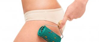
You can do anti-cellulite massages yourself - for this you will need a hard brush or a washcloth for taking a shower, for example. It is preferable to choose products with natural bristles. They perfectly massage the skin, improve blood circulation and promote metabolism at the cellular level. For these purposes, you can also purchase a special massage brush, which is usually used in conjunction with anti-cellulite cream or oil.
At the initial stage, the use of these funds will be sufficient, at later stages they are relevant as auxiliary prevention to consolidate the results.
Treatment options
Treatment requires an integrated approach. Therapy consists of several types of procedures, taking medications, and normalizing lifestyle.
Cosmetical tools
They are effective only with frequent and long-term use. Cosmetics to combat edematous cellulite should have tonic and blood circulation-improving properties. The presence of heating components is considered optimal. Creams for edematous cellulite can be purchased based on reviews from friends or specialists who work with this pathology.
Products should contain extracts of horsetail, hawthorn, horse chestnut, seaweed, clay, caffeine, vitamins and minerals. To enhance the effect, it is recommended to use them during a massage or immediately after a hot shower. Some of them are used directly during hygiene procedures - these can be scrubs and gels for edematous cellulite. After applying cosmetics, you should stay warm, it is best to cover yourself with a blanket or blanket - the warming effect will accelerate tissue regeneration and relieve swelling.
Physical exercise
Performing gymnastics is necessary to increase the tone of the body, eliminate stagnant processes, improve metabolism, and nourish cells with oxygen. Any sport with moderate physical activity is suitable for exercise. Running, swimming, cycling, race walking, and yoga have proven to be the best for relieving swelling.
Procedures in salons
There are several types of procedures that help eliminate the edematous form of the pathology. They must be followed in a course, otherwise they are ineffective. Sometimes the course of therapy is allowed to be repeated after a short period of time. Types of procedures:
- Mesotherapy. Fat burning injections.
- Laser liposuction. Destroying fat deposits using a light beam.
- Endermology. Mechanical impact on tissue using a special apparatus.
- Ultrasound therapy. Activates local blood circulation, corrects the structure of adipose tissue.
- Pressotherapy. Suitable for eliminating edematous cellulite from the extremities.
- Iontophoresis. Exposure to electrical impulses.
- IR therapy. Infrared radiation accelerates blood flow.
- Manual massage. It is carried out using cosmetics.
- Subcision. Indicated in advanced stages. A surgical intervention during which areas with abnormal growth of connective tissue are eliminated.
Courses of some procedures can be carried out simultaneously. This will improve their results and speed up the disappearance of edematous cellulite. Before starting any treatment, you should consult your doctor.
Drinking regime
Drinking at least 2 liters of water per day is an important requirement that must be especially strictly followed during the treatment period. Lack of fluid forces the body to store water, which increases the area of swelling.
As you begin to follow a drinking regime, edematous cellulite may become more noticeable in the first days - this is considered the norm. After their expiration, the volume of edema gradually decreases. This is due to the normalization of metabolism due to the intake of a sufficient amount of fluid into the body, improving blood circulation and lymph flow.
It is impossible to completely remove edematous cellulite by regularly drinking water - this factor is one of the components of the treatment. It is recommended to follow a drinking regime throughout your life to maintain water-salt balance.
Diet
The diet affects the speed of metabolic processes in the body. In order to reduce the appearance of edematous cellulite, you need to follow a special diet. The latter is as follows:
- minimal consumption of salt and sugar;
- the predominance of plant foods in the diet - vegetables, fruits, herbs, nuts, beans;
- refusal of fatty, floury, smoked foods;
- the use of spices and herbs - they are necessary to speed up metabolism;
- choosing lean meat and fish;
- Drink options - herbal teas, natural juices, fruit drinks, still mineral water.
Within a few days after following these rules, a feeling of lightness will appear and the functioning of the gastrointestinal tract will be restored. The reduction in the volume of edematous cellulite will occur gradually.
Folk remedies
Alternative medicine can be useful with recipes for topical use, which accelerate local blood circulation and restore skin elasticity. Masks made from crushed leaves of burdock, dandelion, and algae are suitable for this. They can also be used as a wrapping agent.
You can take herbal decoctions internally to remove excess water from the body. Herbal preparations used to treat kidneys and bladder, and edematous diseases are suitable for this. They may contain thyme, oregano, mint, milk thistle, linden, motherwort, and birch.
Home control methods
Home treatments are less effective than going to a specialist. To achieve the required results, they must be performed daily. Treatment methods:
- Hot baths. High temperatures improve blood flow and relieve swelling.
- Wrap. To carry out the procedure, you can use self-made or purchased products.
- Self-massage. Its effect is enhanced by using special cosmetics for edematous cellulite; it is possible to use rollers, cups and devices.
- Cold and hot shower. The temperature amplitude must be increased gradually. Ideally, very hot water should be replaced by almost ice-cold water every 1-2 minutes.
Before performing an independent massage for edematous cellulite, it is recommended to visit a specialist or watch training videos on the Internet. A few lessons will significantly increase the effectiveness of home therapy.
Additional rules
During treatment, it is necessary to more carefully monitor the condition of your body. In order to facilitate recovery, it is recommended to follow the following rules:
- selection of loose, non-squeezing, breathable clothing;
- wearing compression hosiery – for edematous cellulite against the background of varicose veins;
- avoiding high heels and tight shoes;
- daily home exercises for 15-20 minutes.
Regular adherence to these rules will reduce the likelihood of pathology and vascular diseases of the lower extremities. This occurs due to the normalization of blood circulation and lymph flow.
Types of cellulite
Experts distinguish three main types of cellulite:
- Flaccid (adipose, fatty). The skin is loose and soft, soft tubercles are visible, which can easily change their location. The reason is poor nutrition.
- Hard (fibrous). Very dense lumps on the skin are visually noticeable. The cause of this type of cellulite may be hormonal changes or advanced forms of adipose cellulite.
- Edema (watery). Visually, this cellulite is almost invisible. It appears when pressed. Moreover, it is important that the hole that appears disappears within 2-3 seconds. Only then can you safely say that you have an edematous type of cellulite. It may occur due to hormonal imbalances, poor nutrition, and excessive fluid retention in the tissue.
Varieties
There are several types of edematous cellulite. They differ in their location, degree of distribution, the presence of concomitant pathologies and structure.
Fatty, adipose, or flaccid
This type is diagnosed in obese patients. It is localized on the upper half of the body - arms, stomach, chest. Sometimes it forms on the thighs. Flaccid cellulite is a consequence of an increase in the size of fat cells due to a sedentary lifestyle and poor diet.
The tubercles of adipose cellulite are soft, easily change position, and their palpation does not cause discomfort. The tone of the skin is low, and flabbiness may be detected in some areas.
Hard or fibrous
The result of a violation of the structure and functionality of subcutaneous tissue. The likelihood of hard cellulite depends little on physical activity and nutrition. Its main reason is hormonal imbalance.
Externally, multiple tubercles are detected on the surface of the skin in the absence or insignificant amount of excess weight. The tubercles of fibrous cellulite, unlike edematous ones, are quite hard. Strong pressure causes discomfort and, less commonly, pain.
Hydropic
A consequence of hormonal imbalance, in women it appears when estrogen levels are disturbed. Other possible causes are consumption of large amounts of salt, varicose veins, pregnancy, lipid metabolism failure, and the presence of other types of cellulite.
Edema cellulite is an accumulation of fluid in the subcutaneous fat tissue.
The main distinguishing feature is the preservation of the pit after pressing on the skin for more than 3 seconds. Lumps are large and soft, hard ones are less common. Palpation is not accompanied by unpleasant sensations for the patient.
Salon methods for treating cellulite
Many girls who discover cellulite try to solve the problem on their own. But it should be understood that therapy at home can only be effective in the initial stages. In more advanced cases, the right decision would be to contact a specialized center.
Modern beauty salons offer a huge range of services that are aimed at combating cellulite. But how to choose the most suitable procedure?
Let's highlight the “top 9 salon cellulite treatment methods” in our centers:
- LPG massage is a patented method of body correction and modeling using the 9th generation French devices from LPG Systems - Cellu M6 Endermolab and Cellu M6 Integral. As a result of the impact, dense adipose tissue is kneaded, excess fluid is removed from the body, the appearance of cellulite is smoothed and the condition of the body’s skin is improved.
- LPG Alliance body is a development of the previous method, massage using the 10th generation LPG Systems apparatus. As a result of this procedure, there is not only a decrease in subcutaneous fat, an improvement in skin quality and a reduction in the clinical manifestations of cellulite, but also a pronounced strengthening of the walls of blood vessels, which is reflected in a decrease in the manifestations of varicose veins and vascular networks on the surface of the skin. This procedure is ideal for patients who have vascular problems. The effect is amazing - improving the health of the whole body, slowing down the aging process and modeling a harmonious figure in a short time.
- Pressotherapy is a type of lymphatic drainage, the essence of which is the effect of compressed air on the lymphatic system, which occurs with the help of special corsets and is regulated by a computer. With the help of the procedure, blood supply to tissues is improved, excess fluid and harmful substances are removed. This helps treat tissue affected by cellulite.
- RF lipolysis or radiofrequency therapy has an effect at the level of subcutaneous fat, which improves blood supply, accelerates the process of fat breakdown, hypertrophied fat cells stop putting pressure on the skin - as a result, the condition of the skin improves, cellulite becomes less noticeable.
- Shock wave therapy is the effect of a low-frequency shock wave, which penetrates to a depth of 5 cm and is converted into mechanical energy in tissues, destroying fibrous partitions, stimulating blood circulation, lymph outflow and activating collagen synthesis.
- Biostimulation Futura Pro is a hardware technique for harmonizing body contours, reducing cellulite and strengthening the skin. Special anti-cellulite programs are provided.
- Ozone therapy allows you to reduce the external signs of cellulite at any stage, adjust body volumes and lift the skin.
- Salon wraps (hot, cold, mud-salt, using seaweed, etc.) are ancient techniques based on the application of biologically active substances, followed by the application of bandages.
- Manual classic anti-cellulite massage is a traditional procedure that helps improve blood circulation and metabolic processes in tissues, and improve overall skin tone. This is not only useful, but also very pleasant!
1st stage of cellulite
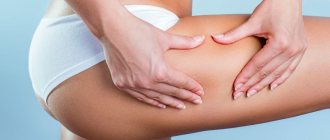
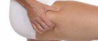
Externally, the first stage of cellulite appears very weakly. Upon examination, loss of skin elasticity is visible in the area of the thighs and buttocks. If you squeeze the skin on the thighs, characteristic signs of cellulite become visible: the skin is very dense, its lumpy structure is clearly visible.
The altered connective tissue lacks oxygen. Toxic decomposition products accumulate there, and the volume of liquid increases. To isolate foreign substances, connective tissue grows, forming a dense jelly-like substance. The original structure of fat cells changes.
Fighting cellulite in the first stage
- The most basic method of fighting at the first stage of cellulite and at the first signs of its manifestation is the restoration of impaired metabolism. This can be achieved with the help of special diets.
- It is also necessary to ensure the outflow of excess fluid from the tissues, which will relieve swelling and normalize cell nutrition. This will help with visiting a sauna, steam baths, and hot wraps.
- Achieving a lasting positive result can be accelerated with daily anti-cellulite self-massage, the use of anti-cellulite cosmetics and physical exercise.