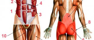
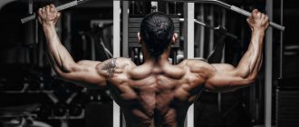
Separation of leg muscles
The lower body has more than 30 muscles. But we are not interested in everyone. Since we are not trying to pass the medical examination. Our goal is to develop the core muscles that will help us achieve outstanding results in the gym. And also, give shape to the lower part of our body. Anatomically, we can divide the leg into 4 parts:
- Pelvis (buttocks)
. These muscles are not exactly part of our leg. But since they are attached to the femur, we take them into account too. And it’s very difficult not to engage your glutes when performing most leg exercises. This muscle group is located on the back side of the body. In the area between the lower back and thigh. Of greatest interest to us are: the gluteus maximus, the gluteus maximus and the gluteus minimus. - Hip. This is the part that almost all athletes want to develop. It is what gives our legs their shape. It starts from the pelvic bone (to which some muscles are attached) and ends at the knee. Its main functions include flexion and extension of the lower leg and thigh. As well as bringing the legs to the body and their rotation (rotation). Conventionally, this group is divided into parts. Anterior, medial (internal) and posterior.
- Shin. These are not large, but very durable muscles that occupy the lower part of the leg. From knee to foot. We are interested in the back group. This includes the triceps and plantaris muscles. All other muscles are responsible for flexion and extension of the fingers. Therefore, we will not consider them.
- Foot.
This is our support. Which is responsible for balance. The muscles located in the foot mainly start from the lower leg. And they are responsible for the movement of fingers. Their flexion and extension. As well as sideways movement relative to each other. And for the turns of the foot. When performing any exercise that requires balancing the body, they are actively involved. Therefore, we will not do separate exercises for these muscles. Still, our task is to devote most of our time to the larger groups.
We can also divide the leg muscles according to the degree of their location:
- Superficial . These are the muscles that are located in plain sight directly under the skin. We can easily touch them with our hands. These muscles give that visual appearance.
- Deep. Located under the surface. And even though they are practically invisible. But they also play a huge role in the formation of legs.
But don't forget one more point. When training our legs, we not only strive to give them one shape or another. We also face the task of making these muscles stronger. This will increase the stability of our body and we will be able to lift heavier weights. And for people involved in strength sports, load progression is one of the key factors.
Features of the structure of the leg muscles
Unlike other muscle groups, the legs have the greatest number of functions. This is the largest collection of muscles that are closely intertwined with each other. Everything is aimed at stabilizing and providing maximum functionality to the three pairs of joints that interact with the muscles of the human legs:
- hip;
- knee;
- ankle
A special role in ensuring human life is played by the muscles of the lower leg: gastrocnemius and soleus. These are powerful pumps that are directly involved in blood circulation. Because of this, calves are often called the “second heart.” Therefore, you need to train your legs not only to increase muscle size or endurance, but also to improve your overall health . Moreover, given the number of muscle fibers and the overall anatomy of the leg muscles, the lower body is designed for heavy workload. This allows you not only to work with heavy weights, receiving a powerful anabolic response, but also to significantly increase your overall endurance (which is more than 50% dependent on the legs).
In sports, the lower body is divided into 4 areas, taking into account their main functions:
- buttocks;
- muscles of the front of the thigh;
- muscles of the back of the thigh;
- calf muscles.
Leg muscles diagram-drawing
For productive training, there is no need to know the names of all leg muscles, taking into account each individual muscle. This is more related to medical topics. However, in order to avoid injury and increase the effectiveness of each exercise, it is necessary to at least generally understand the structure of the muscles of the human legs and the task of each individual area.
Buttocks
From the point of view of the anatomy of the human leg muscles, the gluteal group is considered one of the largest. Moreover, it includes the most massive muscle in the body – the gluteus maximus. The entire area is formed by three gluteal muscles:
- big;
- average;
- small
If you look at the general diagram of the leg muscles, the gluteal structure is designed in such a way that the muscles cover each other in layers, providing mobility and protection to the hip joint. The functions of the gluteal muscles include:
- Straightening the body;
- Taking the leg back;
- Taking your leg to the side.
Gluteal muscles
Front thigh
The entire anterior part is largely represented by a single large muscle, which consists of four heads. From the point of view of the anatomy of the human legs, the muscles and ligaments of the quadriceps are considered one of the strongest. Thanks to this, even beginner athletes can use impressive weights in squats, far exceeding the weight of weights in upper body exercises.
The quadriceps has 4 bundles:
- lateral (outer);
- medial (internal);
- average (intermediate);
- rectus femoris muscle.
The main function of the muscle is to extend the leg at the knee, although the quadriceps is also directly involved in bending forward and flexing the hip.
In addition to the quadriceps, the sartorius muscle also belongs to the front of the thigh. It is considered the longest muscle in the body and runs over the quadriceps.
Leg muscles: anterior group
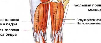
Posterior thigh
Another large muscle area, which is represented by three muscles. If you look at the picture of the anatomy of the leg muscles, the bulk of the back of the thigh is occupied by the biceps muscle (aka “biceps femoris”), which includes two heads:
- long;
- short.
The main function of the biceps muscle is torso extension (works in tandem with the gluteal muscle).
Also included in the back of the thigh are:
- Semimembranosus muscle - responsible for hip extension, flexion and rotation of the lower leg.
- Semitendinosus muscle - performs the same functions as the semimembranosus.
If we evaluate the general purpose of the flexor and extensor muscles of the leg, the main task of the entire posterior surface is flexion of the leg at the knee joint (as well as rotation) . The muscles are also involved in hip abduction and external rotation.
Posterior leg muscles
Adductor muscles
This muscle group is located primarily on the inner thigh. Its main and only function is adduction and supination of the hip. The group includes 5 muscles that form a tightly woven bundle:
- Adductor magnus;
- Long adductor;
- Short adductor;
- Comb;
- Thin.
Leg adductors
This muscle group receives the least attention in fitness, although it plays an important role. If the hip adductors are underdeveloped, they can become a limiting factor in other exercises.
Calf muscles
All muscles of the lower leg are represented by two groups: anterior and posterior. The posterior includes three main and largest muscles , which are closely intertwined into a single bundle:
- Gastrocnemius (aka biceps) - includes the lateral and medial heads. This is the largest muscle below the knee.
- Soleus – located under the gastrocnemius muscle. Athletes who do not know what muscles are in their legs often ignore working the soleus muscle, although it is responsible for the volume of the calves.
- The plantaris is a muscle with long tendons that performs the same functions as the soleus and gastrocnemius. It is considered insignificant and may be completely absent (approximately every tenth person does not have it).
The front part of the lower leg is represented by the tibialis anterior muscle, the main task of which is supination and extension of the foot.
If we evaluate the lower part of the leg muscles taking into account the anatomical description, the main functions of the group include:
- flexion of the ankle and foot;
- supination and extension of the foot;
- rotation of the lower leg (inward).
Leg muscles below the knee
Anatomy of the skeletal bones of the lower limb (leg, pelvis)
Before we start looking at the leg muscles, we'll talk a little about the bones they are attached to. We won’t go too deep, since this is the topic of a completely different article. Below in the picture we can see the skeleton of our lower part.
As you can see, it all starts from the pelvis. Which consists of two iliac bones. The muscles of the pelvis and thigh are attached to their edges, namely the iliac crest. There is also a sacrum, which is attached to the spine. It allows us to keep our body upright. And already at its end, there is a coccyx. Below are two pubic bones. The edges of which serve as the attachment point for the adductor muscles and the back of the thigh. Now let's move on to the leg bones. The largest of them are the femoral ones. At their ends there are two so-called heads, which are attached to the ilium. Next to these heads, there are two protrusions, these are the greater and lesser trochanters. At the lower ends of the femur, there are two epicondyles. Medial (internal) and lateral (external). Also, there are two bones in our lower leg. Tibial and fibular. At the top of the tibia, located are two condyles. Medial and lateral. They serve as places for muscles to attach to them. All the bones of the leg connect to form the knee. There is a small bone there called the kneecap. Better known to everyone as the kneecap. And of course, don’t forget about the bones of the foot. Where are the phalanges of all our fingers located? And the large calcaneus.
This information will be enough to give you an idea. And it will be easier for you to perceive further information.
Causes of pain in leg muscles
Muscle pain (myalgia) in the legs is quite varied and is associated with various types of diseases. One of the most common causes is vascular disease, which is accompanied by swelling and fatigue of the legs, dull pain and ultimately leads to varicose veins.
Also, pain in the leg muscles can be a consequence of problems with the spine. Various pathologies of the spine, such as sciatica or herniated intervertebral discs, can cause radiating pain - pain that radiates to the legs. In this case, there may be no pain in the spine itself. Pain in the leg muscles can also be caused by stereotypical sedentary or standing work, thrombophlebitis (vascular disease), atherosclerosis of the arteries, joint diseases, flat feet, peripheral nerve diseases, infectious bone diseases, prolonged muscle strain and excessive physical activity.
With myositis, pain in the leg muscles is most severe, their intensity increases with movement. Myositis can occur as a complication after various diseases; it can also be caused by injuries, physical overexertion, or muscle damage by parasites. Sometimes myositis occurs as a complication after suffering from the flu.
Fibromyalgia often affects the thigh area near the knee joint. This disease often affects women, and the cause can be both rheumatic diseases and long-term living in damp conditions, psychological and physical stress, injuries and even sleep disorders.
Muscles of the pelvic region
As I said earlier, this includes the gluteal muscles. For the sake of the development of which, a large number of girls in the hall shed a lot of sweat. But this does not mean that men avoid training them. It’s just that their priority is not the round shape, but the strength of these muscles. For example, in powerlifting and weightlifting, the development of the gluteal muscles is given great importance.
Gluteus maximus
It is a very large (hence its name) and flat muscle. The shape is vaguely reminiscent of a rhombus. Its development among all gluteal muscles is a priority. It is located on the surface and covers all the other muscles of this group. At the top it is attached to the posterior surface of the ilium. And also to the lateral edge of the sacrum and coccyx. Directing obliquely downward with its upper bundles, it is woven into the fascia lata (the protective sheath of the muscles). And the lower ones are attached to the gluteal tuberosity of the femur.
Functions:
Extends the hip, abducts it to the side and is responsible for its outward rotation. With fixed legs, rotates the pelvis.
Gluteus medius
This muscle is located immediately under the gluteus maximus. The shape resembles a triangle. Attaches to the outer surface of the ilium wing. Heading down, it turns into a powerful tendon. And is attached to the outer surface of the greater trochanter of the femur.
Functions:
Extends the hip. Participates in hip abduction and is the most powerful muscle that performs this movement. The position of the pelvis stabilizes when we stand on one leg. Also involved in lateral and medial rotation (outward and inward rotation).
Gluteus minimus
This is the smallest and deepest muscle of the three gluteal muscles. The top is covered by the middle one and completely resembles its shape. Just a smaller size. And it is attached in the same places. Above to the outer surface of the iliac wing. And below to the outer surface of the greater trochanter of the femur.
Functions:
Together with the gluteus medius, it moves the leg to the side. Also helps maintain balance when walking. Participates in internal rotation of the hip. That is, in its rotation.
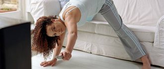
The ORTEKI doctor told readers of the magazine “Liza” how to avoid leg fatigue in the evening
Heaviness, pain, and fatigue in the legs are alarming symptoms that often appear in the evening. They should never be ignored - they indicate that your feet need help and support. What are the causes of such problems and how to avoid them? Sergey Aleksutov, leading doctor - orthopedic traumatologist, chief podiatrist (specialist in foot diseases) of the First professional network of orthopedic salons ORTEKA, tells.
Reason #1. Wrong shoes
Our feet suffer from the wrong shoes, and, as a result, our legs get very tired and “hum” in the evening. Tight shoes put extra strain on the feet even without walking, as it creates strong pressure on the forefoot, which can subsequently lead to deformation of the toes. Every step in high-heeled shoes becomes a real test for the feet: they are exposed to excessive stress, and the joints and spine ultimately take it. Such shoes often cause pain and corns (dry calluses) and provoke the development of flat feet. It is also not recommended to wear shoes with flat soles, since due to insufficient cushioning they do not protect the feet from impacts on the surface.
How to take care of your foot health?
First of all, you need to choose comfortable and safe shoes. A suitable option includes a heel height of 2-4 centimeters, a wide front section and a locking heel. Orthopedic insoles help to take care of the health of your feet; they support the arches of your feet, place them in the correct physiological position, and also reduce stress and strain. In this case, it is best to have your feet scanned by an orthopedic doctor in an orthopedic salon. Based on the characteristics of the feet, the doctor will make individual orthopedic insoles. In comfortable shoes with such insoles, your feet will stop hurting and “humming” by the end of the day.
Reason #2. Lack of movement
An office lifestyle often causes pain, a feeling of heaviness in the legs and swelling. This indicates a circulatory disorder. Blood moves through the veins from bottom to top: from the lower extremities it returns to the heart. The valves of the venous system are responsible for directing the flow of blood. They allow blood to flow to the heart and prevent it from moving in the opposite direction. Most of these venous valves are concentrated in the lower leg. When we move, the muscles in it contract, squeezing blood out of the veins and stimulating its flow to the heart. If the valves do not close enough, reverse blood flow occurs, pressure in the venous system increases, and the walls of the veins become deformed and stretched. Because of this, unpleasant symptoms appear, and varicose veins often begin to develop.
What to do?
Special compression hosiery will help to avoid such problems. It creates the necessary pressure in different parts of the leg so that there is no obstructed blood movement anywhere, and thanks to the effect of distributed compression, it stimulates its flow from the lower extremities to the heart. In addition, for the health of your legs during the working day, try to find time to warm up: light stretching exercises, short walks, at least around the office. Sit often with your legs elevated to promote blood flow. After work, take a contrast shower: alternately turning on slightly warm and cold water improves blood circulation, helping to saturate cells with oxygen.
Reason #3. Huge pressure
The feet take the load from impacts on the surface, protecting the entire musculoskeletal system from it. Due to too intense sports activities, such as running, or work associated with constantly being “on our feet,” our feet become more stressed and more tired in the evening. In addition, due to constant standing, blood circulation in the legs worsens, since blood moves through the veins from bottom to top, overcoming the force of gravity. Without proper attention and care, this lifestyle can cause heaviness and fatigue in the legs, and the development of varicose veins.
How to prevent it?
In cases of increased load on the legs, it is also recommended to wear compression stockings in combination with orthopedic insoles. This will help get rid of unpleasant symptoms. Listen to your body and do not overstrain it with too much physical activity.
Reason #4. Poor preparation
Our legs may simply not physically cope with the load. The muscles and ligaments of the feet need to be strengthened regularly, otherwise regular walking or running will cause fatigue. In addition, weak muscles and ligaments that support the arches of the foot can affect its shock-absorbing function, which can lead to the development of flat feet and heel spurs.
How to train?
Simple, well-known exercises will help: alternating walking on toes and heels, on the inside and outside of the foot, massage of the muscles of the feet, lunges with alternating legs. Such classical workouts stimulate muscle function, strengthen ligaments and joints. They should become your daily feet companions. Of course, you need to try to lead an active lifestyle and play sports. Optimal physical activity on a regular basis in the fitness room will have a beneficial effect on the condition of the whole body. In order to properly develop an exercise program, it is best to seek help from a trainer.
Reason #5. Poor nutrition and its consequences
A lack or, conversely, an excess of energy substances in the diet negatively affects the condition of the entire body, including the health of the legs. Thus, excess weight overloads the muscles and joints of the foot, and also impedes the functioning of the venous system of the legs, which is why they can get sick more often, get tired and even swell in the evening. A person must receive all nutrients in normal quantities: proteins, fats, carbohydrates, vitamins and mineral salts. An imbalance in the consumption of these energy elements can weaken the muscles and ligaments of the musculoskeletal system, particularly the feet. Then even a small load during the day will become a real test for them.
What measures to take?
Proper nutrition is talked about everywhere now, but not everyone adheres to it. First you need to tune in psychologically, realize that this is really important. It’s difficult to change your mind in one day, so you need to start gradually. First, give up excessive consumption of sugar and salt, limit the use of confectionery products, try to exclude processed foods and fast food. Add grain products, vegetables, fruits, dairy and fermented milk products to your diet. Protein foods should not be too fatty. And of course, stick to an active lifestyle to burn extra calories on time.
Attention to yourself
Fatigue, heaviness, pain - this is a cry for help from your legs. Such signs indicate that they need support and care. It is better to react in time, seek help from a doctor, and change your lifestyle to avoid serious health problems in the future.
Be healthy!
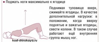
Exercises to train the gluteal muscles
From what was written above, we can notice that these muscles connect the pelvis and thigh. Thus, they become stabilizers for our legs. They also move the leg to the side. But the most important thing is the participation of the gluteus maximus muscle in hip extension. In order to develop them, you need to select exercises where these functions are present. There are several most effective ones:
- Sumo squats. All conditions are met here. Due to the wide stance of the legs, the leg abduction function is activated. When you come into a standing position from a sitting position, the hip extension occurs. And in order not to lose balance, the stabilizing function of these muscles comes into play.
- Romanian cravings. This exercise is aimed at working the buttocks. And it is one of the best for these purposes. The main function it involves is hip extension. Thus, it actively activates the gluteus maximus muscle. And by bending the legs at the knee joint, the biceps femoris muscle is excluded from the work.
- Leg abduction in crossover.
You can move your leg in different directions. Back, focusing on hip extension and working the gluteus maximus. And to the side, engaging the middle and small muscles.
Thigh muscles anterior group
It is this group that forms the thigh from the front. For men, the development of these muscles is of paramount importance. Several main muscles can be identified.
Sartorius
The shape resembles a narrow ribbon. And it is the longest muscle in our body. Its upper end originates from the anterior surface of the ilium. Then it descends obliquely down the front surface of the thigh. And is attached to the tuberosity of the tibia.
Functions:
Participates in flexion of the thigh and lower leg. With fixed hips, the pelvis tilts forward. It also participates in turning the hip inward.
Quadriceps femoris (quadriceps)
This is the largest muscle in our body. From the name it is clear that it consists of 4 heads:
Rectus muscle
The largest of the 4 heads. Located centrally on the front of the thigh. It is this muscle that forms the thickness of the leg. It originates from the lower surface of the ilium. Heading down, it passes into the common tendon. Which is attached to the tibial tuberosity.
Functions:
Due to the fact that the muscle crosses two joints: the hip and knee. She can not only straighten her leg at the knee. But also bend it at the hip.
Important:
When leaning forward during squats, this head is less involved in the work.
Vastus medialis
Located on the anteromedial (inner) surface of the lower thigh. From above it is slightly blocked by the rectus muscle. The shape resembles a drop of water. When well developed, it gives the thigh an expressive shape. From above, a thin tendon is attached to the inner side of the femur. Next, its fibers go down and pass into the broad tendon. With them it partially passes into the common tendon. And the remaining fibers are attached to the inner edge of the patella.
Functions:
Leg extension at the knee joint. Turn the shin inward. Stabilizes the knee, preventing it from falling inward.
Vastus lateralis muscle
Located on the outer part of the front surface of the thigh. Occupying almost the entire area. Slightly covered by the rectus muscle. This head gives shape to the thigh from the outside. Making your legs more massive. In bodybuilding, it is given special importance compared to other strength sports. It originates from the outside of the femur. Heading down, it passes into the common tendon of the 4 heads. And in some bundles it is woven into the lateral (outer) edge of the patella.
Functions:
Leg extension at the knee joint. Rotate the shin outward. Acts as a knee stabilizer. Not letting it fall out. That is, it performs the opposite action of the medial head.
Vastus intermedius muscle
Located on the front of the thigh, immediately under the rectus muscle. It is the weakest among all heads. The upper edge is attached to the anterior surface of the femur. And heading down it passes into the common tendon.
Functions:
Participates in leg extension at the knee joint.
Muscles of the anterior thigh[edit | edit code]
Muscles of the anterior thigh and lower leg
Quadriceps (quadriceps)[edit | edit code]
Quadriceps femoris muscle, m. guadriceps femoris, the most powerful muscle in the human body. It occupies the entire front surface of the thigh. In front it is crossed obliquely by the sartorius muscle
.
Muscles of the anterior thigh
The muscle consists of four heads: rectus femoris, vastus internus, vastus externus and vastus medialis.
. Having different starting points far apart from each other, all the heads converge in the lower part of the thigh and form a common tendon.
Rectus femoris muscle
, m. rectus femoris, bipennate muscle, the longest of the four heads of the quadriceps muscle; located on the front surface of the thigh. The muscle begins as a tendon from the iliac spine and at the acetabulum. Heading down, the muscle expands and reaches the middle of the thigh, and then, gradually narrowing, turns into a powerful tendon. The latter fuses with the base of the patella and, passing along its anterior surface, reaches the tubercle of the tibia, where it ends.
Vastus internus muscle
, m. vastus medialis is a flat, wide, thick muscle. Located on the anteromedial surface of the thigh. Its anterior edge is somewhat covered by the rectus femoris muscle; inside it borders with the medial group of thigh muscles. The vastus internus muscle is partially covered by the oblique sartorius muscle. The muscle bundles, enveloping the anteromedial surface of the femur, are directed obliquely downward and forward. In the lower part of the thigh, the muscle part passes into the tendon part, which joins the rectus tendon.
Vastus externus muscle
, m. vastus lateralis is a flat, wide, thick muscle lying on the anterior outer surface of the thigh. Its lateral surface is partially covered by the tensor fasciae lata muscle; the anterior edge is covered by the rectus femoris muscle. The muscle starts from the trochanter of the femur. The muscle bundles, directed obliquely downward and forward, cover the anterolateral surface of the femur and in the lower part of the thigh pass into the tendon, intertwined with the tendon of the rectus muscle.
Vastus medialis
, m. vastus intermedius - the weakest of the four heads of the quadriceps femoris muscle; has the appearance of a flat, wide, relatively thin muscle. The muscle is located on the front surface of the femur, covered in front by the rectus femoris muscle. The muscle starts from the intertrochanteric line and the anterior surface of the femur within the upper three-quarters. Its bundles are directed vertically downwards and pass into a flat tendon. In the lower thigh, the tendon attaches to the rectus femoris tendon.
Main function:
the quadriceps femoris muscle extends the leg at the knee; in addition, m. The rectus femoris, being a biarticular muscle, takes part in hip flexion and forward tilt of the pelvis.
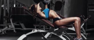
Exercises to train the front of the thigh
As you already understand, this muscle group extends the leg at the knee joint. The most effective exercises that fit these functions include:
- Front squats . Due to the fact that the bar is located in front, we will be forced to tilt it back a little. So as not to drop it. Therefore, most of the load will fall on the quadriceps. The main function involved in this exercise is leg extension.
- Bench press in a machine with narrow legs. Due to the narrow setting, the gluteal muscles are not included in the work. To shift the emphasis to the lateral head, place your feet together. On the medial side, place shoulder width apart.
- Stepping onto the platform. In this exercise, we focus on leg extension. And this is the main function of the quadriceps.
- Leg extension in a sitting machine.
It is a very dangerous exercise. Therefore, you should not use it on an ongoing basis. To avoid damaging your knee joints. From the name it is clear that it meets our requirements. Namely, it extends the leg at the knee joint. When we turn our toes to the side, we load the lateral head more. And inside, medial.
Medial thigh group: adductors
This area is very problematic. And most athletes are developmentally delayed. Because they are not given due attention during training. And without their development, the thigh will not look aesthetically pleasing. Large and developed from the outside. And atrophied from the inside. This group includes the following muscles:
Thin muscle
It has the most superficial location, on the inside of the thigh directly under the skin. The upper edge is attached to the anterior surface of the pubic bone. Then it goes down, around the medial epicondyle of the femur. And is attached to the tibial tuberosity.
Functions:
Takes part in hip adduction and flexion. It also rotates the lower leg inward and takes part in its flexion.
Adductor longus muscle
This is a flat muscle, vaguely shaped like a triangle. Located slightly in front, closer to the quadriceps. It originates from the pubic bone. As it moves downwards, it widens and attaches to the middle part of the femur.
Functions:
Hip adduction, flexion and internal and external rotation.
Adductor brevis muscle
It also has a triangular shape. It is located slightly deeper than the adductus longus. Starts from the front surface of the pubic bone. Next it goes down and is attached just above the middle of the inner surface of the femur.
Functions:
Involved in hip flexion and hip retraction. Stabilizes the pelvis, preventing it from tilting back.
Adductor magnus muscle
This is a wide and thick muscle. She is the strongest of the adducting group. It is located deeper than the long and short muscles, on the inside of the thigh. It originates from a powerful tendon from the bottom of the pubis and ischium. Heading down, it spreads out like a fan onto the wide tendon. And it is attached to the entire inner surface of the femur, and partly to the medial epicondyle.
Functions:
Adducts the hip. Takes part in its bending. Participates in stabilizing the pelvis.
Exercises to train the adductor muscles
As you can see, these muscles are responsible for a stable position. Don't let your pelvis fall back. And of course they bring their legs towards each other. Their development will help improve your posture. Strengthen your inner thighs. Due to this, we can increase strength in many basic exercises. The following are good for their development:
- Plie squats. This is a great basic exercise. Which meets all anatomical functions. By placing the legs wide and turning the feet, we stretch the adductor muscles as much as possible. Also, when coming out of a squat, the thighs are brought towards each other.
- Leg abduction in the simulator. From the name it is clear that the exercise meets our requirements. But when performing it, you must follow the correct technique. There should be no jerky movements. To avoid injuring the adductor muscles.
- Adduction of the leg in a crossover
. Also a common exercise that allows you to work the adductor muscles.
Thigh muscles posterior group
These muscles are antagonists for the anterior group. That is, they perform opposite functions. The front one extends the leg, and the back one bends it. In their training, the main thing is to exclude the gluteal muscles from the work as much as possible. If we don't do this then they will take on most of the load. This group includes:
Biceps muscle (biceps femoris)
Located along the outer (lateral) edge of the back of the thigh. This muscle is the main one in this group. And most of the exercises are aimed at its development. Consists of two heads:
- Long. Which starts from the ischial tuberosity, with a small flat tendon.
- Short. It originates from the inner surface of the lower half of the femur.
Below, these two heads unite into one powerful abdomen. Which passes into a narrow tendon. And bending around the back of the lateral epicondyle of the femur, it is attached to the head of the fibula.
Functions:
Extends the thigh together with the gluteal muscle. Bending the leg at the knee joint. And in this position it can turn it outward.
Semitendinosus muscle
This is a long and thin muscle. It is located closer to the inner (medial) edge of the back of the thigh. Starts from the ischial tuberosity. Heading down, it passes into a thin tendon that wraps around the medial epicondyle of the femur from behind. And is attached to the tuberosity of the tibia.
Functions:
Participates in hip extension and leg flexion at the knee joint.
Semimembranosus muscle
Located along the inner edge of the back of the thigh. Covered below by the semitendinosus muscle. It originates from the ischial tuberosity. Heading down, it goes around the epicondyle of the femur and attaches to the medial condyle of the tibia.
Functions:
Extends the hip. Bends his leg. It also turns the shin inward with the knee joint bent.
Hamstring workout
That is, as we see, exercises should perform two main functions. This is hip extension or leg flexion. We can also use foot rotation in some exercises. When bringing your toes together, the semitransverse muscle is loaded more. When extended to the sides, hamstrings.
- Deadlift on straight legs . This is a basic exercise. Which engages the hip extension function when coming out of a bend. Thus, all the muscles of the posterior group are activated. And since the legs remain straight when tilted. Most of the load will be taken by the back of the thigh.
- Squats with wide legs. Also applies to basic exercises. Involves two functions. Bend the leg at the moment of the squat itself. And hip extension when returning to the starting position. Squats also work all 3 gluteal muscles. And the adductor muscles receive a small share of the load.
- Leg bending in the simulator. Here I think everything is clear. The exercise gives us the opportunity to work the back of the thigh in isolation. By bending the leg at the knee joint.
- Taking the leg back in a crossover. This exercise is mainly performed by girls. It allows you to work all the muscles of the posterior group in isolation.
A little anatomy
Buttocks
Let's start with girls' favorite muscle group - the buttocks.
They consist of three parts - the gluteus minimus, the gluteus medius and the gluteus maximus.
The gluteus minimus and medius muscles are responsible for a toned shape. That is, training aimed at working with these muscle groups will allow you to visually “lift” your buttocks.
The gluteus maximus muscle is the largest muscle in the body and is responsible for shape. If your back naturally extends into your legs, then you should focus on working this particular muscle. Then you can acquire seductive curves.
Anterior thigh
Anterior thigh muscles: quadriceps, pectineus, adductor longus, sartorius and gracilis. All of them are responsible for flexion/extension of the knee and pelvic movements.
Posterior thigh
Hamstring muscles: biceps femoris, semimembranosus, semitendinosus. This muscle group is responsible for the rotational functions of the knee joint and backward movement of the hip.
Shin
Calf muscles: gastrocnemius, soleus, plantar, tibialis anterior. All of them are responsible for rotation of the lower leg and flexion of the foot and ankle joint.
Calf muscles
These muscles are stabilizers for our feet. And is involved in bending the leg at the knee joint. The muscles of the anterior group are responsible for turning the foot, flexing and extending the fingers. But as I said earlier, we will not consider them. We are more interested in the back group. Which includes:
Triceps surae muscle
This is the main muscle that can be trained, and we can influence its shape.
Consists of two muscles:
Calf
lies on the surface and has two powerful heads.
- Medial. It is located closer to the inner part of the back surface of the lower leg. And it is more powerful. Starts from the posterior surface of the medial epicondyle of the femur.
- Lateral. This head, on the contrary, is located closer to the outer part of the lower leg. And it starts from the posterior surface of the lateral epicondyle of the femur.
These two heads are directed downwards and in the middle of the lower leg they are connected into one powerful tendon. The second muscle is the soleus.
This is a flat muscle. Located under the calf. One edge is attached to the head of the fibula. And the other to the inner surface of the tibia. And going down, it connects to the tendon of the gastrocnemius muscle. Which attaches to the heel bone. This tendon is known to many as the Achilles tendon.
Functions:
Participates in bending the leg at the knee joint. Flexes the foot. It is also a stabilizer for the lower leg muscles.
Plantaris muscle
This is a very short muscle that has a long and thin tendon. It originates from the lateral condyle of the femur. It goes down and passes into a narrow tendon that lies between the soleus and gastrocnemius muscles. Attaches to the heel bone.
Functions:
Helps bend the leg at the knee joint. Participates in raising the foot. Tenses the capsule of the knee joint.
Muscles of the lower extremities
Serious pressure is placed on a person’s lower limbs, since it is through them that our movements occur. They can also be considered as support for the upper body. In view of this state of affairs, the muscles of the lower extremities located here are considered one of the strongest and most powerful in humans. Like the muscles of the upper limbs, these are divided into several groups. They are distinguished based on location, as well as functional features. So, there are the muscles of the pelvic girdle; free zone of the lower limb. Despite the visible similarities, a complete analogy between the upper and lower limbs cannot be found, and all because of the serious difference in structure. For example, the scapula and collarbone have fairly serious freedom of movement, but the pelvic girdle is mostly motionless and connects to the spine at the sacroiliac joint.
Thanks to the pelvic muscles, we can move our legs at the hip joint and our spine as well. In turn, this muscle group is divided into two subgroups. As a result, we get the external and internal muscles of the pelvis. The external pelvic muscles include the gluteus maximus, gluteus medius, and minimus, quadratus femoris, and the muscle that is responsible for tensioning the broad connective tissue membrane of the thigh. The same group includes both the superior and inferior gemellus muscles of the lower extremities, including the external obturator muscle. Intrinsic muscles refer to the muscles that form the iliopsoas, obturator internus and piriformis muscles.
The thigh bone is surrounded by thigh muscles. Their doctors divided them into three groups. The result is anterior, medial, that is, middle and posterior. They work to keep the human body upright. But in fact, the main function is considered to be another, namely movement. Just like the pelvic muscles, this group of muscles has evolved throughout human evolution, right up to our species. The thigh muscles have the greatest weight and length. They are capable of developing enormous force, which affects the hip and knee joints.
The muscles of the lower leg are developed much better than others, which is explained by their functionality, namely the upright posture of a person. They originate from bones, intermuscular septa and connective tissue membranes. The calf muscles affect not only the ankle and knee joints, but also the joints of the foot. This group of muscles works on dorsiflexion and plantar extension of our foot. Among other things, their “shoulders” bear the load from the supporting function of the foot and its orientation. All muscles of the lower leg can be divided into subgroups. As a result, we will get anterior, posterior and lateral, that is, side groups. All muscles of the lower extremities involved in extension of the foot belong to the anterior group, while the posterior group refers to the instep supports and flexors of the foot. As for the lateral or lateral group, it includes the flexors and pronators of the foot.
Since a person must always walk straight, the muscles of the foot have a special purpose. Their main functions can rightfully be considered establishing the stability of the foot in different positions, including flexion, counteraction, and abduction of the toes. The muscles of the foot also include the muscles of the dorsum of the foot, the so-called extensors. This also includes the union of the muscles of the plantar part of the foot, which is also called the flexors.
Calf muscle training
Based on what was written above, we can draw a conclusion. That this muscle group receives part of the load in almost any exercise for the legs where we bend them at the knee joint. Plus, they act as a stabilizer for the foot and are also used when rising onto the toes. There are several specialized exercises:
- Calf raises in a special machine. There are a lot of such simulators. There is an option where we perform the exercise standing, sitting and even lying at an angle. Therefore, choose any exercise machine that is in your gym and start raising your toes.
- "Donkey." Refers to a number of rarely seen exercises in modern gyms. But by bodybuilders of the golden era, it was in demand. By tilting the body forward, we stretch the buttocks and the back of the thigh. Therefore, they will not be able to turn on and take the load from the calf muscles.
- Calf raises with dumbbells. This is similar to the first exercise, but instead of a machine, a dumbbell weight is used. For greater stability, take it in one of the hands and perform lifts on each leg in turn.
As you can see, anatomy is an interesting science and its knowledge is very useful. Especially for people who want to develop their body. You don't need to know all the scientific terms. The main thing is to understand what the muscle is roughly attached to. To the pelvis, to the thigh or lower leg and what function it performs. Then you can choose the right exercises.
Good luck to everyone in your training!