Separation of the rectus abdominis muscles, or diastasis, is a common problem affecting mostly women. According to statistics, 70-100% of women develop this pathology in the third trimester of pregnancy. Normally, after childbirth, the structures should return to their original position, but in approximately 30% of cases this does not happen. A gap remains between the muscle tissues, which causes a bulging, flabby abdomen and provokes more serious consequences. That is why it is important to know how to determine diastasis of the abdominal muscles at home, which doctor can help, and what treatment methods modern medicine offers.
What is diastasis
To understand this concept, it is necessary to briefly outline the structural features of the abdominal muscles. In the center of the abdomen is the rectus muscle. It begins in the lower abdomen and extends to the pubic bone. Consists of two sections that are connected in the center by loose fabric. This connective tissue is called the linea alba.
As a result of prolonged and strong tension of muscle structures, intra-abdominal pressure increases. Weak tissues of the white line cannot withstand the load and begin to diverge. At the same time, the distance between the sections of the rectus muscle increases, they diverge to the sides and move several centimeters away from each other. A kind of furrow is formed, and when tense, the stomach protrudes unnaturally. Pathological discrepancy can form under the navel, above it, or have a mixed form.
The pathology should not be confused with a hernia of the white line or with an umbilical protrusion. It does not threaten to infringe on internal organs. But at the same time, even at an early stage it represents an aesthetic problem, since diastasis of the rectus abdominis muscle is much more often determined in women than in men.
Depending on the degree of discrepancy and the size of the formed distance, diastasis is divided into three degrees:
- first - the distance between sections is from 2.5 to 5 cm;
- second - the discrepancy reaches a gap of 5-8 cm;
- third - the size of the furrow between the sections exceeds 8 cm.
This classification allows you to understand how to determine the degree of muscle diastasis even at home.
Pathology can also affect other muscle structures of the anterior abdominal wall. Depending on what surrounding tissues are involved in the process, four types of diastasis are distinguished:
- A - classic form, in which only the rectus muscle diverges;
- B - loss of tone in the inferolateral muscle sections;
- C - appearance of expansion in the area of the xiphoid process, ribs;
- D - curvature of the waist line.
Service
How is diastasis sutured?
There are several fundamentally different approaches to surgical correction of diastasis of the rectus abdominis muscles.
Most often, diastasis is sutured during abdominoplasty by our colleagues - plastic surgeons. Unfortunately, in the vast majority of cases they are limited to simple suturing of the white line (the so-called “plication”), sometimes with two rows of sutures.
Simple plication with “regular” absorbable suture material carries a risk of recurrence of diastasis (not to mention hernia) in 40% of cases, according to a study conducted by Van Uchelen et al [1]. Of course, such results cannot suit us.
Plastic surgery with local tissues without the use of a mesh implant is quite acceptable for small diastasis, up to 40 mm wide, and a hernia with a defect of up to 10 mm. This has been proven and recommended in the clinical guidelines of the European Hernia Society (EHS). When performing plastic surgery of the anterior abdominal wall, we use in our practice the latest generation non-absorbable suture material, “anchor” type (V-loc PBT), which, in our opinion, is much more reliable than traditionally used options, and this more than compensates for its high cost.
Good results are demonstrated by suturing the diastasis using tangential sutures, which we use in some cases.
Fig. 1. Tangential sutures during open suturing of diastasis of the rectus muscles make it possible to make the relief of the anterior abdominal wall flatter [2]
Diastasis of more than 40 mm or the presence of a hernia with a defect of more than 10 mm is an indication for the use of a mesh implant (“mesh”).
The mesh implant serves to strengthen the sutured white line and/or hernia defect. It makes no sense to simply close her diastasis, which I have observed with surprise several times among my colleagues - with this option, the protrusion of the white line after the operation occurs along with the mesh, and there is no cosmetic result of this operation.
In principle, the implant can be placed in different layers of the anterior abdominal wall:
We use all options, choosing the optimal one for each patient in a specific clinical situation. Except perhaps for option B, which has long proven its ineffectiveness.
We rarely use the location of the implant in the abdominal cavity (intra-abdominal). The fact is that polypropylene (or polyester), which the implant itself is made of, should not come into contact with the abdominal organs - it causes a massive adhesive process, leading to adhesive intestinal obstruction and other life-threatening complications. Implants for intra-abdominal plastic surgery have a special anti-adhesion coating on one side, which significantly increases the cost of the implant, but is not always able to “protect” the organs. And if recently these were isolated cases, today more and more often voices are heard among herniologist surgeons about abandoning this technique. I gave it up several years ago, after twice operating on patients (from other clinics, and even countries) with complications. Yes, this is not much, but why expose the patient to even a minimal risk of complications that can be avoided?
Rice. 2 Options for placing a mesh implant for hernia and diastasis repair: A – above the aponeurosis (onlay), B – sewing into the edge of the defect (inlay), C – in the axillary space, D – in the preperitoneal space (underlay preperitoneal), E – in the abdominal cavity (underlay intraperitoneal) [2].
The choice of the optimal layer for implant placement depends on many factors: from the presence and width of aponeurosis defects and the degree of diastasis to the body mass index and skin condition of the patient. Each option has its own advantages and disadvantages; we choose it together with the patient during consultation.
Suturing of diastasis
Diastasis is a condition in which the distance between the rectus abdominis muscles increases. There are two methods of performing the operation - tension and non-tension. The rehabilitation period is relatively easy. At first there is swelling, hematomas, and pain. The first ones gradually go away on their own, and the pain is relieved with a drug prescribed by the doctor. Cost of the operation:
from 170,000 rubles Make an appointment with a doctor
More about the operation
Causes of abdominal muscle diastasis
The main reason for the discrepancy is the excessively high pressure that occurs in the abdominal cavity. At the same time, the prerequisites for increased intra-abdominal fluid are of different nature. These include:
- Loss of muscle elasticity as a result of rapid weight loss.
- Excessive physical activity.
- Dysplasia is the abnormal development of tissues and organs. In addition to diastasis, it is accompanied by many additional manifestations: hernia, varicose veins, hemorrhoids, etc.
- Pregnancy. As a result of hormonal changes, collagen production is reduced, tissues lose elasticity and become loose. At the same time, the enlarged uterus significantly increases pressure on weakened muscle structures and the linea alba.
In 60% of cases, diastasis in women is associated with pregnancy. Pathology begins to develop in the middle of the second trimester. It is at this time that the muscles are stretched under the influence of increasing abdominal pressure. Normally, after childbirth, the uterus restores its previous size, and the width of the white line returns to its normal value of up to 2 cm.
However, in many cases, the recovery process is complicated by concomitant factors that prevent the tissues from returning to their previous position. These factors include:
- mature age of the woman in labor;
- excess weight before and during pregnancy;
- the fruit is too large;
- number of previous pregnancies and births;
- type of pregnancy (one fetus or several);
- complications during pregnancy;
- too rapid return to active physical activity after childbirth.
Diastasis can also occur in children, especially premature babies. The key prerequisite for the development of pathology is the failure of the child’s muscles and tendons. In this case, most often the defect is eliminated on its own during the first year of the baby’s life. During this period of time, the muscles acquire tone, the ligaments become strong. Only children with Down syndrome are at risk of maintaining the discrepancy.
In only 1.5% of cases, diastasis is diagnosed in men. The basic prerequisites are the same as for women: obesity, dysplasia, sudden weight loss. Also, men are more likely to be overly involved in strength training, which not only causes hernias and varicose veins, but also provokes the formation of diastasis.
Signs of abdominal muscle diastasis
For a long time, the pathology has an asymptomatic course. Manifestations increase as the discrepancy develops and complications appear. And if in women the signs of diastasis of the rectus abdominis muscles are maximally manifested after pregnancy and childbirth, then in men - only when the pathology is already in an advanced state.
The main and most noticeable manifestation of the pathology is a rounded vertical protrusion of the abdomen. If you deliberately tense your abdominal muscles, a groove between the right and left halves of the body becomes visible. At the same time, in men, even with intense strength training, the abs are not sufficiently worked out and do not acquire the desired relief.
If the disease progresses, the pathological structure of muscle tissue and disturbances in muscle function become the cause of other, more dangerous characteristic manifestations. There are:
- pain in the spine, lower back;
- posture disorders;
- increased fatigue;
- dysfunction of the gastrointestinal tract, which is accompanied by heartburn, belching, pain, constipation, flatulence.
At the third stage of the pathology, dangerous complications are possible, which manifest themselves in the form of:
- ptosis, prolapse of internal organs;
- intestinal obstruction;
- urinary incontinence;
- renal colic;
- feeling of heaviness in the legs while walking;
- muscle atrophy in the abdominal area.
How can a vacuum be dangerous?
Anastasia:
Vacuum is a kind of massage of internal organs. And therefore, the main danger of this exercise is that it can provoke an exacerbation of existing diseases of the organs and biliary tract or create additional cramps during menstruation. In addition, performing this exercise significantly stimulates an increase in blood pressure, so if you have problems with the cardiovascular system, it is worth consulting your doctor.
It should be borne in mind that a vacuum also stimulates blood circulation, so it can provoke the spread of viral diseases or malignant tumors throughout the body and worsen the condition.
Photo: unsplash.com/@szabolcs_taking_pictures
Like any other exercise, vacuum has contraindications and is not suitable for everyone who wants to correct their posture or acquire a slender waist. If you are not sure that the exercise is right for you, or you experience pain while doing it, you should consult a specialist.
Anastasia:
The vacuum cannot be used during menstruation, pregnancy, diseases of the uterus, acute infectious diseases of the abdominal and pelvic organs, exacerbations of chronic digestive diseases, thromboembolic disease, as well as malignant tumors of any location.
You should not perform a vacuum during the rehabilitation period after operations in the abdomen and pelvis. Also, you should not perform a vacuum on a full stomach. At least one and a half hours should pass since the last meal. Ideally, perform on an empty stomach in the morning.
How does a doctor determine the presence of diastasis of the rectus abdominis muscle?
If any of the listed symptoms appear, as well as if there are predisposing factors such as recent pregnancy and childbirth or increased physical activity, you should consult a doctor for examination. You can’t delay your visit; it’s better to see a specialist for prevention than to wait until dangerous complications appear.
Many people do not know which doctor determines the presence of diastasis of the rectus abdominis muscles. You need to contact a surgeon. In most cases, an experienced specialist will be able to determine the pathology using only palpation examination.
For diagnosis, the patient lies on his back, slightly bends his knees and rests his feet on the surface. After this, the surgeon asks you to tighten your abdominal muscles. At the same time, you need to raise your shoulder blades and head. The doctor feels the abdomen, measures the width of the discrepancy, and determines the presence and stage of the pathology. In this case, pronounced diastasis of the third degree is noticeable even in a standing position.
In some cases, the study of the width of the white line is complicated by excess body weight. The doctor refers such patients for an ultrasound examination. This procedure is also prescribed if there is a suspicion of the development of complications: hernial protrusions, displacement of internal organs. In rare cases, radiography or computed tomography is necessary.
Possible negative consequences
Abdominal exercises are allowed only if there are no restrictions. If you perform the task during menstruation, negative consequences may arise in the future in the form of changes in the cycle.
Comment! You can perform the complex while sitting on a chair, kneeling or resting on your hands.
Menstrual irregularities are another possible harm to the female body. To avoid negative effects, you can start vacuuming the abdomen during menstruation already at the end of the period of bleeding.
How to determine the presence of abdominal muscle diastasis yourself
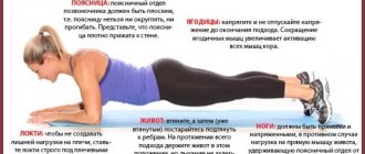
Diastasis can be determined at home. A simple test will help you do this, during which you need to measure the distance between the edges of the rectus muscle.
To do this, you need to lie on your back on a hard surface, bend your knees. Place one hand under your head and the other on the white line, just above your navel. Then you should relax and raise your chest. In this case, you need to feel the discrepancy between the muscles with your fingertips. The gap is most pronounced between the navel and the xiphoid process. If the discrepancy is greater than the width of two fingers, this should alert you and be a reason to contact a specialist.
This technique is as similar as possible to the approach that a surgeon uses for diagnosis. This is the easiest way to determine diastasis at home without the help of a doctor and instrumental examinations.
How to remove diastasis
The treatment regimen for the pathology depends on the stage of its development and the degree of manifestation. At the first stage, it is enough to follow the doctor’s recommendations, which will help strengthen the muscles of the abdominal wall and reduce their discrepancy:
- reduce weight to normal levels;
- maintain a balanced diet with limited consumption of fatty and sweet foods;
- adhere to the daily fluid intake;
- wear a bandage that supports the stomach;
- undergo massage and other physical procedures;
- do swimming, yoga, Pilates or physical therapy.
Physical therapy should be carried out under the supervision of a specialist. He creates a program with the correct load level. Particular attention is paid to the deep transverse and oblique abdominal muscles, which, with an appropriate level of tone, reduce the degree of stretching of the linea alba.
A set of therapeutic exercises is useful even for pregnant women. With its help, you can prevent the development of pathology and speed up the process of postpartum recovery of the body.
Such training takes place without straining the abdominal muscles. It is also not recommended to do exercises while standing, including on your knees or elbows. Such loads are permissible only after restoration of the normal width of the lumen.
In later stages, the discrepancy can no longer be corrected with exercise or massage. Surgical intervention is necessary to return the muscle corset to its place and relieve complications and symptoms. It could be:
- Tensionplasty using patient tissue. The surgeon removes excess connective tissue and stitches the edges of the muscles together. This method is the least preferable, since relapses occur quite often after it.
- Tension plastic using a prosthesis. It involves the same removal of excess tissue and suturing of muscle layers, but is accompanied by additional strengthening using a mesh polypropylene prosthesis.
- Tension-free plastic surgery with installation of a prosthesis. An endoprosthesis is inserted under the stretched area, which serves as a barrier and replaces the weakened structures.
- Combined approach. It involves a combination of tension and non-tension plastic surgery.
The specialist selects the surgical method taking into account the characteristics and degree of development of the pathology, as well as individual factors and the patient’s health status. Full recovery after the intervention occurs within 1 to 3 months. At this time, it is necessary to avoid excess tension, adhere to a diet and wear a special bandage that relieves stress from the operated muscle structures.
Why you can’t do a vacuum during menstruation
The main reason why it is not recommended to do exercise during menstruation is increased bleeding. The mechanism of action on the reproductive system looks like this:
- the girl performs the indicated exercises;
- tensing the abdominal muscles, the uterus also tenses;
- When the uterus is tense, bleeding increases.
Increased uterine bleeding provokes harm to the body, and subsequently cycle disorders and amenorrhea may develop. Gynecologists who claim that you can do a abdominal vacuum during menstruation mean the 5-6th day of the menstrual cycle. During this period, there is less bleeding, so you can safely start working on your body.
Prevention of diastasis
To reduce the risk of developing pathology, it is extremely important to systematically follow preventive recommendations. To do this you need:
- adhere to a proper balanced diet;
- lead an active lifestyle, walk more;
- strengthen all muscle groups, especially the muscle structures of the abdomen and lumbar region;
- eliminate excessive physical activity;
- do not lift weights, especially for women;
- strengthen the diaphragm;
- monitor your weight and prevent obesity.
During pregnancy, you need to use special oils, creams, ointments that increase the elasticity and firmness of tissues. After childbirth, it is necessary to carefully monitor the condition of the press in order to detect pathology in a timely manner and, if necessary, seek help from a doctor before complications develop. Treatment of the initial stages of discrepancy is successful and does not require excessive effort from the patient.