Diastasis is an increase in the distance between two sections of the rectus abdominis muscle. The separation occurs at the abdominal suture (Linea Alba and Linea Semilunaris) of the midline connective tissue in the anterior abdomen.
This phenomenon can occur after pregnancy, with rapid weight gain or loss, prolonged and significant physical activity, prolonged cough or regular constipation. The formation of diastasis is promoted by congenital weakness of connective tissue (dysplasia).
Diastasis is often combined with hernias of the anterior abdominal wall of various locations, varicose veins of the lower extremities, flat feet, hemorrhoids, blurred vision and scoliosis.
Strong and prolonged tension of the abdominal muscles leads to an increase in intra-abdominal pressure, and weakness of the linea alba leads to the fact that the muscles, under the influence of the increased intra-abdominal pressure created by them, begin to diverge to the sides. The so-called “vicious circle” closes when muscle tension causes a further increase in diastasis.
During pregnancy, under the influence of the hormone relaxin, the ligaments are stretched and the muscles of the anterior abdominal wall are weakened so that the uterus, and with it the abdomen, can expand with the growth of the fetus. After childbirth, the muscles gradually recover, but the tendons may remain in their previous state and create the appearance of a belly protruding forward.
In addition to a cosmetic defect, diastasis can be dangerous for a woman’s health, since a number of mechanisms that ensure the proper functioning of intra-abdominal structures, abdominal and back muscles are disrupted, and, as a result, pelvic discomfort and prolapse of internal organs may occur .
66% of women with diastasis of the rectus abdominis muscles note the development of urinary incontinence when coughing or sneezing, and discomfort during sexual activity. Diastasis and various pelvic disorders usually go hand in hand.
| 100% of women have some degree of diastasis recti in the third trimester of pregnancy, this means that physiological diastasis cannot be avoided, it is important to monitor the recovery afterwards. |
It is important to note that for many women, rectus discrepancy persists for 6-8 weeks after giving birth, and this discrepancy may remain unchanged for up to a year after giving birth. At the same time, a discrepancy of up to 2 cm is considered a physiological norm.
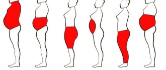
There are 3 degrees of diastasis depending on the size of the discrepancy of the rectus abdominis muscles:
1st degree - 2-5 cm; 2nd degree - 5-7 cm; Grade 3 - more than 7 cm.
There are three types of diastasis depending on the location of the discrepancy:
- Above the navel;
- Below the navel;
- Mixed type (presence of muscle divergence both above and below the navel).
In 2009, a group of scientists conducted a study - using ultrasound methods, the width of the white line was measured in 150 primiparous women aged 20-45 years.
| It has been found that the average width of the linea alba can vary greatly, from 7 mm at the xiphoid process to 13 mm above the umbilicus and 8 mm below the umbilicus. |
Causes
Most often, the problem worries women; diastasis of the rectus abdominis muscles after childbirth appears in almost every fourth woman in labor. Miniature mothers of two or more children who have a delicate constitution are especially susceptible to excessive stretching of the aponeurosis of the rectus muscles. Women who were involved in sports before pregnancy and have a thick build are least at risk. Diastasis of the rectus abdominis muscles in men often appears due to an incorrectly selected set of strength exercises, and in young athletes due to coaching errors. The constitutional characteristics of children can provoke the onset of the disease in the absence of an individual approach to the choice of exercises. The so-called dysplastic children have a congenital weakness of the connective tissue, which also makes up the bridge (aponeurosis) between the muscles. Diastasis of the rectus abdominis muscles in a child: with existing weakness of the connective tissue, with constant loads leading to increased intra-abdominal pressure, an abnormal divergence of the superficial rectus abdominis muscles is formed. Young dysplastic athletes can be identified by a number of characteristics:
- detect MAPSS syndrome - minor cardiac anomalies;
- valgus shape of the lower extremities (X-shaped legs);
- flat feet with valgus deformity (most of the load falls on the inner edge of the foot);
- frequent ankle subluxations.
In pregnant women, a temporary state of thinning, “softening” of tissue along the midline of the abdomen results from:
- increased intra-abdominal pressure due to a growing uterus;
- Hormonal changes contribute, which contribute to greater compliance of the ligamentous apparatus of the pelvic bones (for the safe passage of the fetus through the birth canal).
Repeated births with diastasis of the rectus abdominis muscles lead to an even greater intermuscular defect. Often, women, in an attempt to regain their previous shape, resort to rigorous strength training, but an incorrectly selected set of exercises for diastasis of the rectus abdominis muscles after childbirth leads to great disappointment - the discrepancy of the muscles can increase due to increased intra-abdominal pressure during exercise. And the annoying bulge of the abdomen will not disappear, because it is impossible to train the aponeurosis (tissue between the muscles) - there are no muscle fibers there and it will not decrease after physical exercise. exercises.
Signs, symptoms and causes.
Diastasis at the initial stage does not manifest itself with any typical symptoms. In a person, a keel-shaped protrusion of the abdominal wall appears in the middle of the abdomen, and when the press is tense, a semicircular depression appears in the center of the abdomen.
At a later stage, a patient with diastasis may experience the following unpleasant symptoms.
Him:
- severe discomfort appears, pain in the epigastrium, in the navel, pelvis, and lower back;
- difficulties arise while moving.
Remember: diastasis is a very insidious disease. Because of it, the functioning of the gastrointestinal tract can quickly be disrupted. In this case, the patient suffers from urinary incontinence, which occurs when he tenses the peritoneum, sneezes, or coughs. Additionally, he may experience congestion in the blood vessels of the legs.
Thus, if a person has one of the symptoms described above, then he immediately needs to see a doctor and undergo a full examination.
The patient can also identify diastasis on his own.
All he needs is:
- Lie down on a flat surface.
- Bend your lower limbs at the knees.
- Tighten your abdominal muscles.
In addition, the patient can perform a slightly different test.
He needs:
- Lie on your back, bend your knees, place your feet on the floor.
- Raise one hand and place it under your head.
- Raise your other hand, place its fingers in the middle of the abdomen, i.e. to the place where a person’s abs are located.
- Lightly press your fingers on your stomach.
- Raise your upper body, twist your torso to one side.
- Identify the presence of diastasis and its width.
In this position, a person can see a strong protrusion located in the center of the abdomen, which has fairly even contours. And when feeling this area and the navel, the patient may identify a small depression located between the muscles.
Note: people who are overweight will not be able to detect diastasis at home.
Additionally, the patient will have to undergo instrumental research methods.
He will need to do:
- Ultrasound of the abdominal cavity. It can reveal thinning and stretching of the linea alba, an increase in the space between the vertical muscles. Also, using ultrasound, you can determine whether the patient has hernias and excessive prolapse of the internal organs of the peritoneum.
Most often, people wonder why they have diastasis. It usually occurs with increased intra-abdominal pressure, weakness of the connective and muscle tissue of the peritoneum.
Do not forget that diastasis usually appears in the fairer sex after childbirth. It is during the period of carrying a child that the fairer sex experiences strong intra-abdominal pressure, which negatively affects the abdominal muscles.
In addition, diastasis can appear in the fairer sex:
- with multiple pregnancies, excess weight;
- bearing large fruit;
- with polyhydramnios, loose skin of the anterior abdominal wall.
Why do pregnant women suffer from this disease?
It's simple.
They have:
- intra-abdominal pressure increases. Its increase is caused by a sharp increase in the size of the uterus;
- Serious hormonal changes occur in the body. Such changes lead to softening of the ligamentous apparatus of the pelvic bones. Thanks to its softening, the baby passes better through the birth canal.
Moreover, with repeated births, diastasis appears again in women, but in a more serious form. Many women, after giving birth again, go on a strict diet and resort to serious physical exercise. But an incorrectly selected set of exercises leads to even greater divergence of the abdominal muscles. Because of this, the bulge on the stomach becomes more defined. Remember: it is almost impossible to get rid of severe diastasis with the help of physical activity. In this place a person has no muscle fibers, so it is almost impossible for people with diastasis to pump up their abs.
In addition, diastasis very often torments young children.
Most often it appears in children:
- with weak connective tissue;
- exposed to constant physical activity, during which intra-abdominal pressure increases, an abnormal divergence of the rectus abdominis muscles occurs.
Children with diastasis are very easy to identify.
They have:
- there are slight changes in the functioning of the heart;
- Valgus curvature of the lower extremities occurs. Typically, children with diastasis have their legs crossed;
- flat feet and valgus deformity of the lower extremities occur, where most of the load falls on the inner part of the foot;
- Frequent ankle subluxations occur.
Thus, if a person has diastasis, then the following typical symptoms bother him.
He experiences:
- pain, discomfort in the abdomen, which intensifies when performing physical activity;
- dyspeptic disorders. He often suffers from constipation, bloating, nausea;
- such ailments as splanchoptosis. In which the organs located in the abdominal cavity descend and shift excessively and unpleasant sensations arise;
- pain while walking or in a standing position.
Also, in a patient with diastasis, the muscles of the anterior abdominal wall often atrophy.
Types, methods and principles of treatment of diastasis
Not only visually, but also externally, the patient can independently identify diastasis of the abdominal muscles. So even he can determine its degree.
1 finger=1 cm.
Normally, the width of the discrepancy between the muscles should be no more than 2 cm.
Conventionally, doctors distinguish 3 stages of diastasis of the abdominal muscles.
This is diastasis:
- First degree. With it, the patient feels a muscle discrepancy of 3-7 cm.
- Second degree. The patient may personally notice a muscle discrepancy of 7-10 cm.
- Third degree. In this case, the patient may notice a discrepancy in the muscles of 10 cm or more. Also, with third-degree diastasis, the patient’s midline constantly protrudes, and the structure of the abdomen changes.
In the first days after childbirth, women may develop a diastasis of 2.5 cm or more. After 1-1.5 months, this disease goes away on its own.
Do not forget that the operation is not performed on all patients with diastasis of the rectus abdominis muscles.
For example, it is prohibited if the patient has:
- dangerous illnesses. This may be a disease of the kidneys, blood, liver, diabetes;
- infections, acute inflammation.
Symptoms
Usually the manifestations of the disease are not pronounced; patients are more concerned about sagging abdomen and changes in figure. With a significant degree of diastasis, symptoms may appear:
- Pain, discomfort in the abdomen, aggravated by physical activity;
- Dyspeptic symptoms - constipation, bloating, nausea;
- Splanchoptosis – prolapse, displacement of the abdominal organs with corresponding symptoms;
- Discomfort while walking, while standing;
- Atrophy of the muscles of the anterior abdominal wall.
What is diastasis recti
This is an increase in the space between the right and left abdominal muscles compared to normal.
Normally, these muscles are connected to each other by the linea alba. Doctors call this line the tendon aponeurosis. This is a light plate formed from dense collagen fibers. When the interstitial tissue is stretched, a person experiences various cosmetic and medical complications. Conventionally, doctors believe that diastasis is a divergence of the vertical muscles of the anterior abdominal wall, located between the pubic bone and the ribs. The tendon membrane connecting the longitudinal muscles is located in the center of the abdomen. During diastasis, it becomes thinner, weakens, and ceases to hold the abdominal muscles in the anatomically correct position. It is because of this that they move apart.
Muscle discrepancy is not only associated with cosmetic defects. Such citizens have poor muscle function; the muscles are not able to restrain intra-abdominal pressure.
With a strong deviation from the norm, a person develops a hernia of the white line, and the navel protrudes excessively. At the same time, his internal organs rest against the skin and suffer greatly from any mechanical impact.
In addition, diastasis also leads to the appearance of splanchnoptosis in humans. With it, a person’s muscle tissue weakens, and the internal organs become severely depressed. Such patients often suffer from bloating, constipation, and nausea. They also experience increased heart rate and frequent dizziness.
Usually this disease worries representatives of the fairer sex. Very rarely, diastasis appears in men. Its appearance is associated with the work activity of men. This disease also often occurs in men who do not watch their weight and often eat at fast food restaurants.
Diastasis leads to increased load on the spinal column. Therefore, such citizens often suffer from back pain and distorted posture.
How to determine diastasis rectus abdominis muscles
How to check diastasis recti at home? There is a simple method. In many cases, diastasis “catches” the eye even without additional examination - right along the midline on the abdomen when coughing or straining, a longitudinal ridge forms, sometimes up to several tens of centimeters long. If diastasis is not so pronounced, you can perform a simple test:
- You need to lie on your back, bend your knees and rest your feet on the floor;
- Raise one hand and place it under your head;
- Place the fingers of the second hand along the midline of the abdomen at waist level, above and below the navel, lightly pressing them on the abdominal wall;
- Lift your upper body while twisting your torso;
- When lifting, determine the diastasis that appears and its width. Usually it is felt as a certain “dip” of the fingers deep into the abdomen, between the muscles.
By the distance felt by the fingers, one can approximately determine the diastasis of the rectus abdominis muscles and its degree. One finger is taken as 1 cm (approximately). Normally, the width of the aponeurosis between the rectus muscles (they are the most superficial and powerful) is no more than 2 cm.
1st degree of diastasis - if a discrepancy of more than 3 cm is detected - up to 7 cm.
Grade 2 is diagnosed with a defect of 7-10 cm.
Stage 3 of the process - the distance between the rectus abdominis muscles is more than 10 cm, constant protrusion in the midline, change in the configuration of the abdomen.
In women, several weeks after childbirth, a diastasis of 2.5 cm or even more can be detected. In most cases, the situation changes for the better after a month and a half. To increase the chances of returning to your previous figure, it is necessary to adjust your diet in the very early postpartum period and begin specialized exercises to strengthen the abdominal muscles (this will also have a beneficial effect on uterine contractility and the prevention of late postpartum hemorrhage).
Not all strength exercises on the anterior abdominal wall can improve the situation, quite the contrary. The exercise therapy complex for diastasis of the rectus abdominis muscles excludes exercises with lifting and twisting of the torso, including on the wall bars.
Exercises to strengthen the abdominal muscles
Inclusion of oblique abdominal muscles, lateral bends, various variations of plank pose and specially adapted training for our students, taking into account increasing load from minimal to intense.
You can choose any 3-5 exercises and start with 10 repetitions on each side.
Side plank scissors, knee on the floor
Oblique abdominal muscles, back muscles, lateral thigh muscles, arm muscles.
Starting position (IP): lie on your side, support on your elbow, elbow strictly under your shoulder, supporting leg bent at the knee, back straight.
We lift the pelvis off the floor, the body is tense, there is one straight line from the shoulder to the knee. Raise the straight left leg up, then lower it to the floor and lower the pelvis into the IP.
Side plank scissors
Oblique abdominal muscles, back muscles, lateral thigh muscles, arm muscles.
IP: lie on your side, support on your elbow, elbow strictly under your shoulder, legs straight, back straight.
As you exhale, lift your pelvis off the floor and lift your upper leg as high as possible. With an inhalation, we lower it and return to the starting position. IMPORTANT: do not lean to the side, the body is as tense as possible.
Side fold
Oblique abdominal muscles.
IP: lying on your side, the lower arm lies along the body, the other hand behind the head (elbow looking to the side). Legs are straightened, in the air, parallel to the floor (but do not lie on it!).
On the count of “one”, with an exhalation, we perform twisting to the side, trying not to just bend our legs at the knees, but to raise them as high as possible, reaching with our elbows towards our knees. The supporting arm bends at the elbow during twisting. On the count of “two” we return to IP. IMPORTANT: keep your feet on the floor throughout the entire exercise. We don’t lean forward or backward.
Dynamic side plank with forearm (knee on floor)
Oblique abdominal muscles.
IP: side plank from the forearm, the elbow is clearly under the shoulder, the supporting leg is bent at the knee 90 degrees and lies on the floor, the back is straight, the pelvis is twisted, the body is one straight line.
Slowly raise and lower your pelvis, tensing your abdominal muscles. IMPORTANT: the back is always straight, raise the pelvis as high as possible, try not to touch the floor at the lowest point, stop the pelvis 1-2 cm from the floor.
What's not allowed?
Prohibited exercises for diastasis recti
The “correct” exercises for diastasis of the rectus abdominis muscles are the Pilates system. Such exercises will help strengthen the deep transverse muscle, which duplicates the stretched aponeurosis. The transversus muscle will help maintain adequate intra-abdominal pressure and keep the abdominal organs in their normal position. Diastasis of the rectus abdominis muscles in men - the choice of exercises is the same as for women.
Pranayama for diastasis
Breathing exercises will help you regain control of your abdominal muscles and strengthen your inner wall. It is most convenient to perform pranayama while sitting with your legs crossed or on your heels. It is important that you feel comfortable keeping your back straight.
Full yogic breathing
Place one palm on your stomach. The other is on the chest so that the thumb and index finger touch the collarbone. Begin to inhale deeply from bottom to top: first, the stomach protrudes forward, then the ribs expand, and finally the collarbones rise. Hold your breath for 1-2 seconds and begin to exhale slowly in the same order: stomach, chest, collarbones.
At first, you can inhale for 4 seconds and exhale for 4 seconds. Over time, you can gradually lengthen both inhalation and exhalation. Practice full yogic breathing for 3-4 minutes.
Kapalbhati
Inhale calmly, and as you exhale, contract your abdominal muscles, pushing out the air. Inhalation then occurs voluntarily. Breathe in this way at a rhythm of one inhale-exhale per second.
At first, it is enough to perform 3 circles of 20 inhalations and exhalations. After some time, you can gradually increase the number of inhalations and exhalations.
Treatment of diastasis of the rectus abdominis muscles
The situation with the appearance of divergence of the abdominal muscles is not corrected spontaneously.
For degrees 1-2 of diastasis, it is recommended to carry out adequate training, breathing exercises, and adhere to a diet, and then you can expect an improvement in the condition of the muscle wall and a reduction in aesthetic defects. If diastasis has developed as a result of chronic lung diseases or digestive disorders, it is necessary to treat them as the root cause. Taping of diastasis of the rectus abdominis muscles is a modern and convenient auxiliary method for correcting the defect; you can learn how to apply tapes quite quickly, the procedure is painless and does not cause inconvenience.
Considering that the aponeurosis does not contain muscle fibers, with weakness of the deep muscles (including the transverse one), with the 3rd degree of diastasis, the logical and practically the only choice for eliminating the defect is surgical treatment.
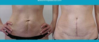
Surgery for diastasis of the rectus abdominis muscles - photo. The results of endoscopic surgery are without scars.
Our center performs the most popular and modern operations for diastasis of the rectus abdominis muscles, using endoscopic methods in accordance with international standards for the quality of medical care. According to indications, treatment is supplemented with abdominoplasty and liposuction to improve the aesthetic effect of the operation.
As a result of surgical intervention, the patient’s appearance radically changes for the better, and traces of the operation are almost invisible.
Depending on the condition of the tissues in the area of diastasis, the degree of protrusion and stretching of the abdomen, and the patient’s weight, the following can be applied:
- Transumbilical video-assisted ventral alloabdominoplasty. The most gentle operation, without scars and scars - through a minimal incision in the navel area, only 4 cm long. Using a laparoscope and under visual control (the image from the endoscope camera is displayed on the screen, enlarged several times), a graft is placed under the anterior abdominal wall and fixed with sutures to underlying muscles. This transplant is made entirely of inert (non-aggressive) materials to the body and does not cause allergic or immune reactions. Its mesh structure allows it to quickly assimilate with the patient's tissues. Such an operation may be accompanied by hernioplasty (if there is an umbilical hernia and diastasis of the rectus abdominis muscles). There are no relapses of diastasis; with endoscopic alloabdominoplasty, the percentage of complications does not exceed 1%, and the postoperative period is minimized.
- The laparoscopic method of suturing diastasis of the rectus abdominis muscles using the plication method is a surgical treatment option when the aponeurosis defect is eliminated by bringing the muscles together and stitching them together, while the peritoneum is sutured together with the aponeurosis along the entire length of the diastasis. The operation is minimally invasive - instruments are inserted into the operating area through two 3-4 cm incisions (near the navel and in the bikini area). Depending on the need, it can be supplemented with the use of an inert graft using the IPOM method. This kind of “patch” is used if the discrepancy between the muscles is large or a hernia of the white line is detected.
- Traditional operations with open access (incision) are surgical procedures, in some cases applicable and selected by the doctor according to indications (initial tissue condition and degree of diastasis, presence of concomitant pathologies, etc.). But nowadays they are resorted to less and less often, since after such treatment of diastasis of the rectus abdominis muscles, after surgery, cosmetic defects (scars) remain and relapses are likely. And the postoperative period is quite long and painful.
There are several options for such “academic” operations:
- Operation with extraperitoneal access according to Champion. Through an incision in the skin and subcutaneous fat, the surgeon gains access to the rectus muscles and aponeurosis, the muscles are sutured along the edge and pulled towards each other, the excess aponeurosis is immersed inside.
- Surgery for diastasis of the rectus abdominis muscles with intraperitoneal access according to Voskresensky. During the operation, the abdominal cavity is opened and a kind of duplication of the connection between the left and right rectus muscles is carried out by placing a double suture, first on the inner surface of the muscles facing the abdominal cavity, and then on the outer one. In this case, excess aponeurosis and peritoneum are removed.
- The Kockerling method of surgery is distinguished by the use of a mesh graft for plastic surgery of the anterior abdominal wall with access through a traditional incision.
- The Troitsky method with intraperitoneal access is applicable if diastasis is accompanied by a postoperative hernia of the white line of the abdomen.
For diastasis of the rectus abdominis muscles, surgery; its price will depend on the chosen method of surgical intervention, the method of anesthesia and the subsequent rehabilitation period (the volume of rehabilitation and resuscitation measures performed). If you have diastasis of the rectus abdominis muscles, you can find out the real price of treatment after consulting a surgeon.
Bandhas for diastasis
Bandhas, or locks, are contractions of certain muscles that are used both during the practice of asanas and pranayamas, and separately. I have previously written in detail about what locks are in yoga and why they are needed.
There are three locks in yoga. To combat diastasis, we will need two of them. It is best to perform both locks while sitting cross-legged or on your heels.
Mula bandha (root lock)
Sit cross-legged or on your heels. Tighten your pelvic floor muscles, contract the perineum and anus. You may not be entirely sure how to control these muscles at first. For convenience, you can imagine that you want to connect the pubic bone and tailbone - you will feel how the necessary muscles “tighten”. Then imagine that you want to connect your sit bones - again, the right muscles will be activated.
Hold Mula Bandha for 10 seconds, then take a 5-second break. Perform the root lock for 2-3 minutes. You can gradually increase the time you hold the lock.
Postoperative period
After endoscopic interventions, the patient remains in the hospital for no more than 2 days, and after 14 days he can gradually return to his usual physical activities. Patients report feeling good already on the first day after surgery, because in most cases epidural anesthesia is used, which eliminates the difficult recovery period after general anesthesia. A bandage for diastasis of the rectus abdominis muscles is recommended for everyone without fail for a period of 1 month.
During operations with traditional access, the time frame for returning to a normal lifestyle is significantly longer; lifting weights of more than 10 kilograms and physical exercise for about 3 months are not recommended. The biggest negative point for patients in such cases is quite pronounced pain in the postoperative area in the first days and the appearance of a scar 10 cm or more in length. But these operations in some cases are the best choice in terms of prognosis.
Our clinic uses all modern methods of surgical elimination of diastasis of the rectus abdominis muscles. Operations can be supplemented with abdominoplasty and liposuction, which is especially important in the treatment of diastasis of the rectus abdominis muscles after childbirth.
Exercises to correct diastasis
It is important to note that the approach to correcting diastasis through exercise should be gradual and regular . Our system involves gradually engaging the deep core muscles to ensure an even load. As a result, this approach leads to the closure or significant reduction of diastasis.
| You can perform a set of exercises to correct diastasis in grades 1 and 2, in the absence of hernias and no earlier than 6 months after birth by cesarean section and 1 month after natural birth (start from the first stage of exercises). |
If you have grade 3 diastasis, you should stop at the first stage of the program until it reaches at least grade 2.
The #sekta diastasis rehabilitation program is conditionally divided into two stages:
- the first stage is the formation of a physical activity regime;
- the second stage is the transition to more intense exercises.
Work on correcting diastasis begins with correcting or strengthening posture, walking, joint exercises, normalizing diet, breathing exercises, strengthening the transverse abdominal muscle and pelvic floor muscles.
Pre-operative reminder for patients with diastasis
Pre-operation memo for patients
- click on the image to download.
Before surgery
before hospitalization
Before hospitalization, you will need:
- come to an appointment with the surgeon to receive a reminder and instructions
- undergo all necessary examinations before hospitalization
- bring compression stockings of class 1 compression, if necessary (the doctor decides) - class 2
on the eve of hospitalization
- make sure that all original examinations necessary for hospitalization are available
- Check with your doctor for the exact time of arrival at the emergency department
on the day of hospitalization (the day before surgery)
- in the morning before leaving for the hospital, take a shower (you can use antibacterial agents)
- undergo a hospitalization procedure, examination by a surgeon and an anesthesiologist in the hospital
- the evening before surgery - a light dinner until 20:00, then only liquids are allowed
- before bed - shower
On the day of surgery
- immediately after waking up, without getting out of bed, put on compression stockings
- In the morning before surgery you cannot eat or drink. An exception is taking a sip of water to wash down the medicine.
- honey. the staff will help with shaving the surgical area and guide you to the operating room
- in the operating room, the anesthesiologist will once again explain the anesthesia procedure and actions after the operation
- In preparation for the operation, you will have a venous catheter and, if necessary, an epidural analgesia system installed.
- you will come to in the operating room or intensive care unit
- If an epidural catheter is present, it will be removed in the intensive care unit
- after that you will be transferred to a ward
Exercises for diastasis
The complex is simple and short - you can include it in your exercise program and/or do it during the day as an exercise break.
Marjariasana (Cat Pose)
• Get on all fours, palms under your shoulders, knees under your pelvic bones. • Slightly pull in your stomach, smoothing out the lumbar deflection - your back is straight. • Fingers spread to the sides, middle fingers pointing forward. Support is not only on the base of the palm, but also on the upper phalanges of the fingers. • With an inhalation, bend over (see the main photo for the article above), opening the chest and as if trying to connect the crown and tailbone through the top. Emphasis on deflection in the thoracic region, not in the lumbar region. Don’t let your stomach fall out as you inhale—it’s always in a slight tone. • As you exhale, round your back, try to “connect” the crown and tailbone through the bottom. We draw in the stomach well, the navel tends to the spine. The emphasis is on the lumbar region, we try to round it as much as possible.
Take your time, stretch out the breathing cycle. It is optimal to breathe full yogic breathing in 4 counts (4 seconds inhale and 4 seconds exhale) or slower. Perform the movement 8-10 times.
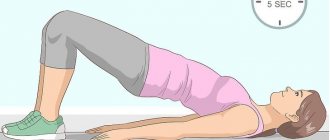
Setu Bandha Sarvangasana (Half Bridge)
• Lie on your back with your arms extended along your body, palms down. • Bend your knees and place your feet close to your buttocks. • As you inhale, slowly begin to move your pelvis and your back up behind it. When you reach your shoulder blades, tuck your shoulders in and push your chest toward the ceiling. Continue to lean on your shoulders and feet. • Your feet should be under your knees—that is, your shins form a 90-degree angle with the floor. • Lengthen your neck, point your chin slightly toward you, and move your shoulders away from your ears. • Tighten your pelvic floor muscles, contract your buttocks, and push your pelvis up even harder. • Hold this position for 20-30 seconds. Breathe, slightly stretching your usual breathing cycle. • Exhale, vertebra by vertebra, roll your back as smoothly as possible on the mat, and lower your pelvis last.
Rest for a few seconds and repeat the exercise 4-5 more times.
Twists
• Lie on your back. Feet on the floor, knees bent and looking up. The arms are extended to the sides, forming one straight line, palms facing up. • Exhale, lower your knees to the right and turn your head to the left. • Inhale and return to center. • Exhale and twist to the other side.
Perform 4-5 twists in each direction. At the end, you can stay static on each side for 20-30 seconds.
Pavana Muktasana (Wind Release Pose)
• Lying on your back, pull your knees to your chest and hug them with your arms. • Relax your stomach, breathe calmly and evenly.
Stay in this position for 30-40 seconds.
How do you know if you have diastasis?
Lie on your back, bend your knees, place your feet on the floor. Relax your head and shoulders and place your fingers (palm facing your face) directly above your navel.
Lift your head and neck very slowly up and press with your fingertips in the area above the navel. If you feel a dip inward, it means diastasis. Try not to lift your shoulders off the floor. Repeat the test several times in different places.
The stretch is measured by the width of the fingers. Most often, this discrepancy is 1-2 fingers wide, if more, then do not panic.
More important in this case is the tension and its lack in the area of the white line. When the muscles contract, there should be tension and resistance when you press your fingers into the area of the white line. If this is not the case, then the connective tissue needs to be restored.
What exercises and movements are best to avoid?
Twisting, squats, torso twists, planks, leg lifts from a lying position and holding them suspended are not recommended for diastasis.
Exercises to work the transverse press and to align and strengthen the core and pelvic floor muscles can be performed at any stage of diastasis development. If you do a lot of crunches or pump your abs, then most likely the sprain condition will worsen and the stomach will take on the characteristic “dome” shape. This is an example when the muscles were worked hard, but in the wrong direction. You won't be able to completely restore them to their previous position or pump them up, but you can restore strength, stability, and improve the appearance of your belly.
Do I need to wear a brace, bandage or elastic bandages?
The postpartum bandage has been used at all times. Abdominal support during pregnancy and immediately after it helps in some cases, you just need to wear it correctly. This will give stable support to the abdominals and lower back.
But even if you tighten the muscles tightly using a clamp, it does not guarantee that the white line will recover and remain in the same place. That is why you should not rely on a retainer or elastic bandages to return everything to its place, otherwise you will have to wear it constantly and for a very long time.
Remember that diastasis occurs due to excessive stress within the abdominal cavity and pelvis. This is pressure that the body cannot withstand, although it should. A brace and brace will not fix the problem; they will simply bring the two pieces back together after they have come apart, but will not restore the connective tissue between them. First you need to restore balance to the whole body and use the core muscles to remove diastasis.